Dermatitis Herpetiformis: Pathogenesis and clinical findings
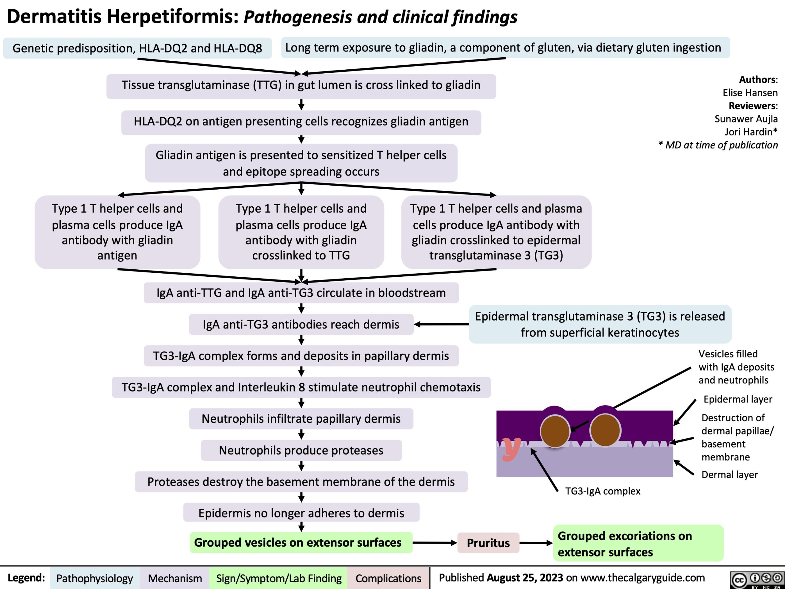
Genetic predisposition, HLA-DQ2 and HLA-DQ8 Long term exposure to gliadin, a component of gluten, via dietary gluten ingestion
Tissue transglutaminase (TTG) in gut lumen is cross linked to gliadin
HLA-DQ2 on antigen presenting cells recognizes gliadin antigen
Gliadin antigen is presented to sensitized T helper cells and epitope spreading occurs
Authors: Elise Hansen Reviewers: Sunawer Aujla Jori Hardin* * MD at time of publication
Type 1 T helper cells and plasma cells produce IgA
antibody with gliadin antigen
Type 1 T helper cells and plasma cells produce IgA
antibody with gliadin crosslinked to TTG
Type 1 T helper cells and plasma cells produce IgA antibody with gliadin crosslinked to epidermal transglutaminase 3 (TG3)
IgA anti-TTG and IgA anti-TG3 circulate in bloodstream
IgA anti-TG3 antibodies reach dermis
TG3-IgA complex forms and deposits in papillary dermis TG3-IgA complex and Interleukin 8 stimulate neutrophil chemotaxis Neutrophils infiltrate papillary dermis
Neutrophils produce proteases
Proteases destroy the basement membrane of the dermis Epidermis no longer adheres to dermis
Epidermal transglutaminase 3 (TG3) is released from superficial keratinocytes
Vesicles filled with IgA deposits and neutrophils
Epidermal layer
Destruction of dermal papillae/ basement membrane
Dermal layer
Y
Grouped vesicles on extensor surfaces
Pruritus
TG3-IgA complex
Grouped excoriations on extensor surfaces
Legend:
Pathophysiology
Mechanism
Sign/Symptom/Lab Finding
Complications
Published August 25, 2023 on www.thecalgaryguide.com