Superficial Thickness Burns: Pathogenesis and clinical findings
Author: Amanda Eslinger Elise Hansen Illustrator: Amanda Eslinger Reviewers: Alexander Arnold Sunawer Aujla Yan Yu* Duncan Nickerson* * MD at time of publication
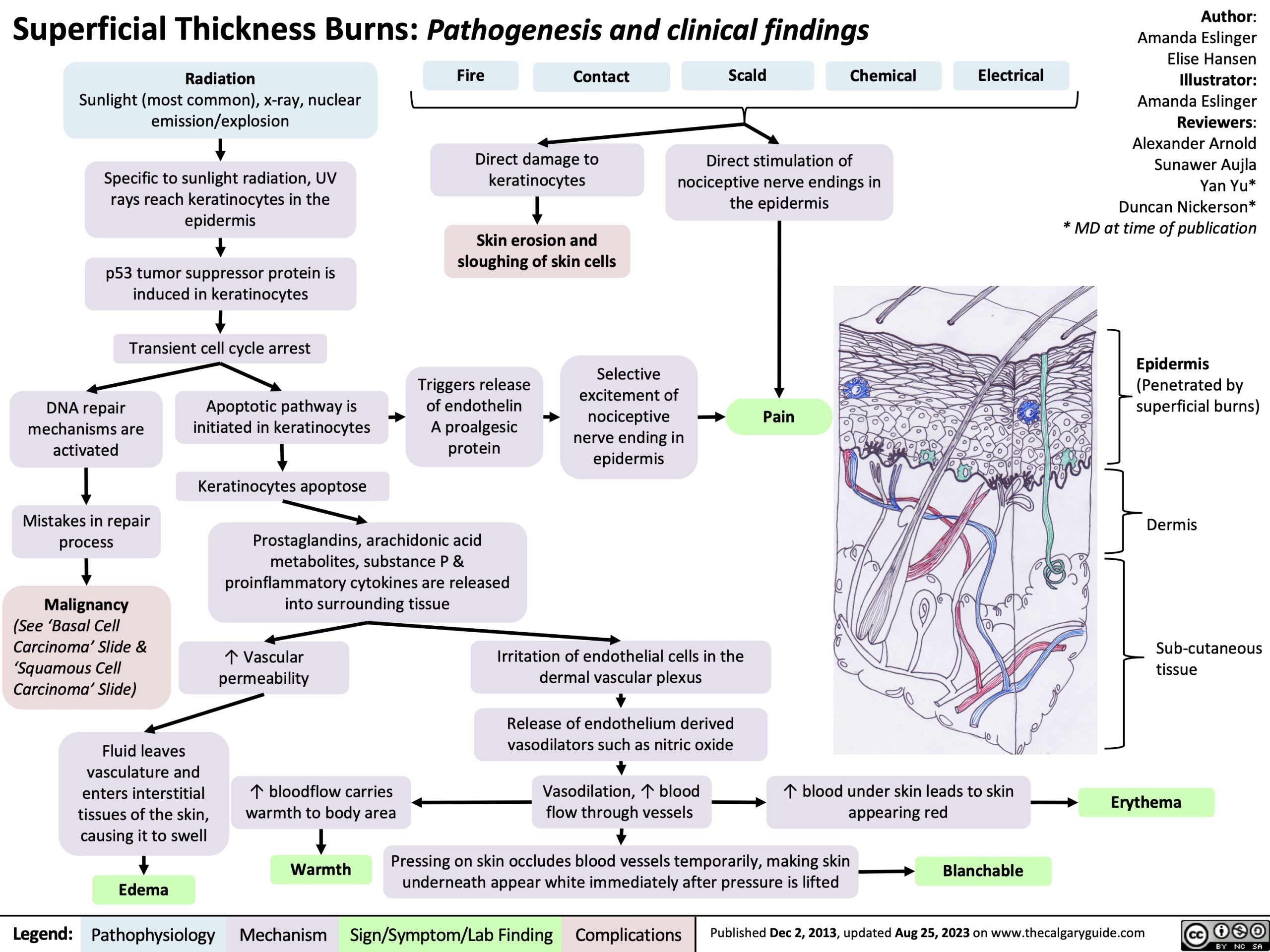
Epidermis
(Penetrated by superficial burns)
Dermis
Sub-cutaneous tissue
Erythema
Radiation
Sunlight (most common), x-ray, nuclear emission/explosion
Specific to sunlight radiation, UV rays reach keratinocytes in the epidermis
p53 tumor suppressor protein is induced in keratinocytes
Transient cell cycle arrest Apoptotic pathway is
Fire
Contact
Scald
Chemical
Electrical
Direct damage to keratinocytes
Skin erosion and sloughing of skin cells
Direct stimulation of nociceptive nerve endings in the epidermis
DNA repair mechanisms are activated
Mistakes in repair process
Malignancy
(See ‘Basal Cell Carcinoma’ Slide & ‘Squamous Cell Carcinoma’ Slide)
Fluid leaves vasculature and enters interstitial tissues of the skin, causing it to swell
Edema
Triggers release of endothelin A proalgesic protein
Selective excitement of nociceptive nerve ending in epidermis
Pain
initiated in keratinocytes
Keratinocytes apoptose
Prostaglandins, arachidonic acid metabolites, substance P & proinflammatory cytokines are released into surrounding tissue
↑ Vascular permeability
↑ bloodflow carries warmth to body area
Irritation of endothelial cells in the dermal vascular plexus
Release of endothelium derived vasodilators such as nitric oxide
Vasodilation, ↑ blood flow through vessels
↑ blood under skin leads to skin appearing red
Warmth Pressing on skin occludes blood vessels temporarily, making skin Blanchable underneath appear white immediately after pressure is lifted
Legend:
Pathophysiology
Mechanism
Sign/Symptom/Lab Finding
Complications
Published Dec 2, 2013, updated Aug 25, 2023 on www.thecalgaryguide.com