Acute and Chronic Subdural Hematoma on CT: Pathogenesis and findings
Brain shrinkage with age or alcohol misuse
Head trauma
Congenital or acquired coagulopathy (i.e. anticoagulant use)
Other (i.e. brain mass)
Stretching & tearing of bridging cortical veins that cross from the cortex to dural sinuses
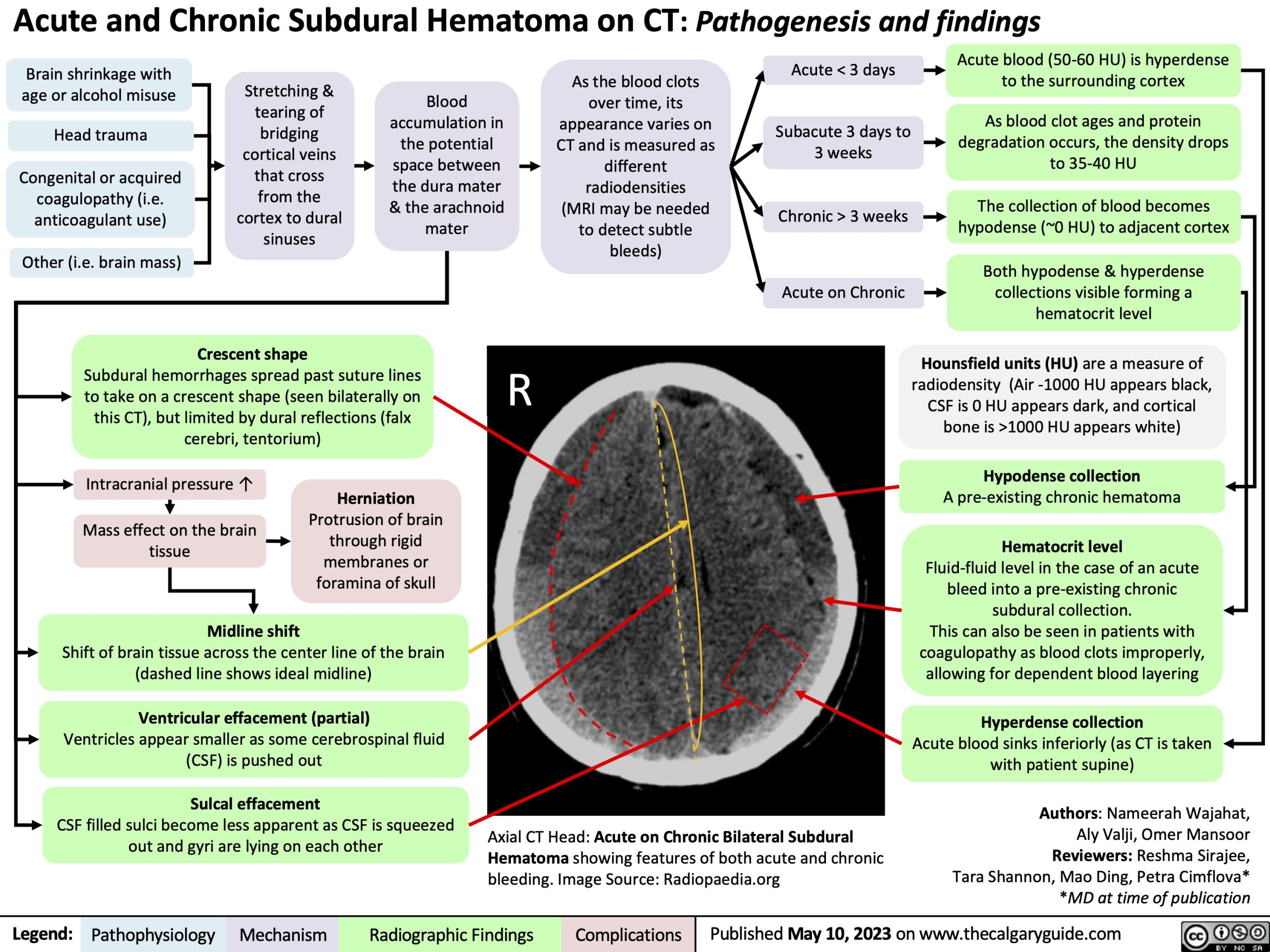
Crescent shape
Blood accumulation in the potential space between the dura mater & the arachnoid mater
As the blood clots over time, its appearance varies on CT and is measured as different radiodensities (MRI may be needed to detect subtle bleeds)
Acute < 3 days
Subacute 3 days to 3 weeks
Chronic > 3 weeks Acute on Chronic
Acute blood (50-60 HU) is hyperdense to the surrounding cortex
As blood clot ages and protein degradation occurs, the density drops to 35-40 HU
The collection of blood becomes hypodense (~0 HU) to adjacent cortex
Both hypodense & hyperdense collections visible forming a hematocrit level
Hounsfield units (HU) are a measure of radiodensity (Air -1000 HU appears black, CSF is 0 HU appears dark, and cortical bone is >1000 HU appears white)
Hypodense collection
A pre-existing chronic hematoma
Hematocrit level
Fluid-fluid level in the case of an acute bleed into a pre-existing chronic subdural collection.
This can also be seen in patients with coagulopathy as blood clots improperly, allowing for dependent blood layering
Hyperdense collection
Acute blood sinks inferiorly (as CT is taken with patient supine)
Authors: Nameerah Wajahat, Aly Valji, Omer Mansoor Reviewers: Reshma Sirajee, Tara Shannon, Mao Ding, Petra Cimflova* *MD at time of publication
Subdural hemorrhages spread past suture lines to take on a crescent shape (seen bilaterally on this CT), but limited by dural reflections (falx cerebri, tentorium)
R
Intracranial pressure ↑
Mass effect on the brain tissue
Midline shift
Shift of brain tissue across the center line of the brain (dashed line shows ideal midline)
Ventricular effacement (partial)
Ventricles appear smaller as some cerebrospinal fluid (CSF) is pushed out
Sulcal effacement
CSF filled sulci become less apparent as CSF is squeezed out and gyri are lying on each other
Herniation
Protrusion of brain through rigid membranes or foramina of skull
Axial CT Head: Acute on Chronic Bilateral Subdural Hematoma showing features of both acute and chronic bleeding. Image Source: Radiopaedia.org
Legend:
Pathophysiology
Mechanism
Radiographic Findings
Complications
Published May 10, 2023 on www.thecalgaryguide.com