Small Bowel Obstruction: Findings on X-Ray
Authors: Evan Allarie Shelley Spaner* Reviewers: Davis Maclean Yan Yu* * MD at time of publication
See Calgary Guide – Mechanical Bowel Obstruction and Ileus: Pathogenesis and clinical findings
Bowel contents cannot pass the obstruction
Buildup of bowel contents (gas, fluid) proximal to the obstruction
Gas rises above the fluid
If exclusively or mostly accumulation of fluid (and not gas) occurs
Any small amounts of gas/air present (not enough to create an
air-fluid level) will rise and become trapped in valvulae conniventes (small bowel folds)
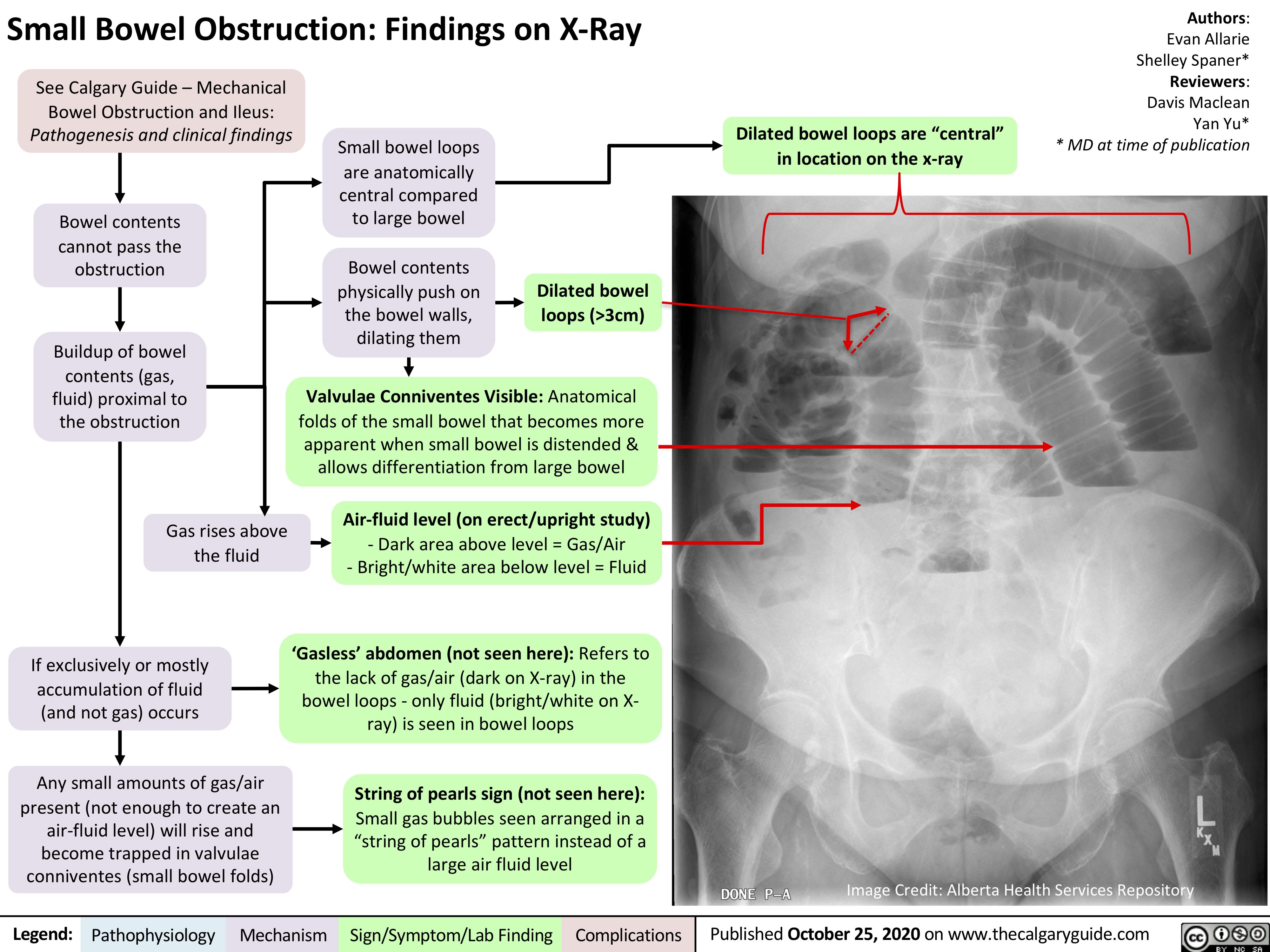
Small bowel loops are anatomically
central compared to large bowel
Bowel contents physically push on the bowel walls, dilating them
Dilated bowel loops are “central” in location on the x-ray
Dilated bowel loops (>3cm)
Valvulae Conniventes Visible: Anatomical folds of the small bowel that becomes more
apparent when small bowel is distended & allows differentiation from large bowel
Air-fluid level (on erect/upright study)
– Dark area above level = Gas/Air
– Bright/white area below level = Fluid
‘Gasless’ abdomen (not seen here): Refers to the lack of gas/air (dark on X-ray) in the bowel loops – only fluid (bright/white on X- ray) is seen in bowel loops
String of pearls sign (not seen here):
Small gas bubbles seen arranged in a “string of pearls” pattern instead of a large air fluid level
Image Credit: Alberta Health Services Repository
Legend:
Pathophysiology
Mechanism
Sign/Symptom/Lab Finding
Complications
Published October 25, 2020 on www.thecalgaryguide.com