Rotator Cuff Disease: X-ray and ultrasound features
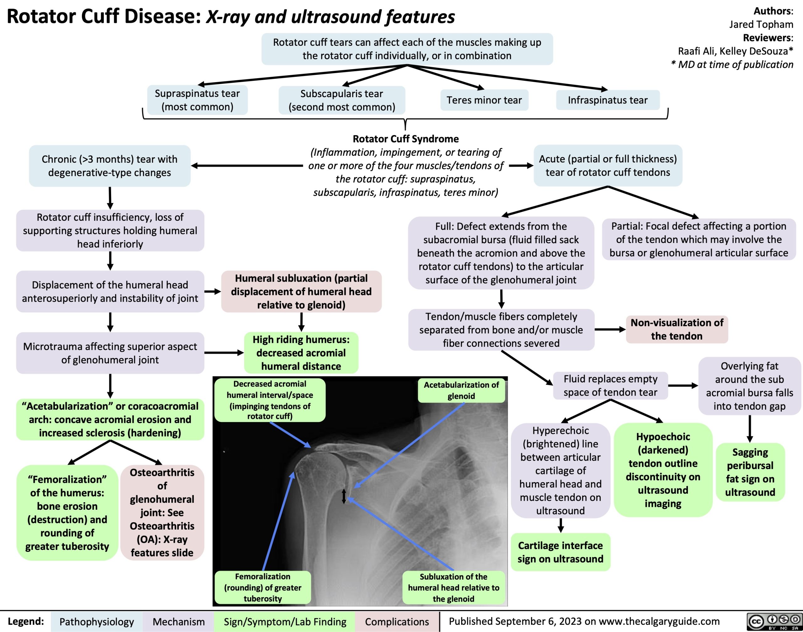
Rotator cuff tears can affect each of the muscles making up
the rotator cuff individually, or in combination
Authors: Jared Topham Reviewers: Raafi Ali, Kelley DeSouza* * MD at time of publication
Supraspinatus tear (most common)
Chronic (>3 months) tear with degenerative-type changes
Rotator cuff insufficiency, loss of supporting structures holding humeral head inferiorly
Displacement of the humeral head anterosuperiorly and instability of joint
Microtrauma affecting superior aspect of glenohumeral joint
“Acetabularization” or coracoacromial arch: concave acromial erosion and increased sclerosis (hardening)
Subscapularis tear (second most common)
Teres minor tear
Infraspinatus tear
Rotator Cuff Syndrome
(Inflammation, impingement, or tearing of one or more of the four muscles/tendons of the rotator cuff: supraspinatus, subscapularis, infraspinatus, teres minor)
Acute (partial or full thickness) tear of rotator cuff tendons
Humeral subluxation (partial displacement of humeral head relative to glenoid)
High riding humerus: decreased acromial humeral distance
Decreased acromial humeral interval/space
(impinging tendons of rotator cuff)
Full: Defect extends from the subacromial bursa (fluid filled sack beneath the acromion and above the rotator cuff tendons) to the articular surface of the glenohumeral joint
Tendon/muscle fibers completely separated from bone and/or muscle fiber connections severed
Partial: Focal defect affecting a portion of the tendon which may involve the bursa or glenohumeral articular surface
Non-visualization of the tendon
Acetabularization of glenoid
Fluid replaces empty space of tendon tear
Overlying fat around the sub acromial bursa falls into tendon gap
Sagging peribursal fat sign on ultrasound
“Femoralization” of the humerus: bone erosion (destruction) and rounding of greater tuberosity
Osteoarthritis of glenohumeral joint: See Osteoarthritis (OA): X-ray features slide
Hyperechoic (brightened) line between articular cartilage of humeral head and muscle tendon on ultrasound
Cartilage interface sign on ultrasound
Hypoechoic (darkened) tendon outline discontinuity on ultrasound imaging
Femoralization (rounding) of greater tuberosity
Subluxation of the humeral head relative to the glenoid
Legend:
Pathophysiology
Mechanism
Sign/Symptom/Lab Finding
Complications
Published September 6, 2023 on www.thecalgaryguide.com