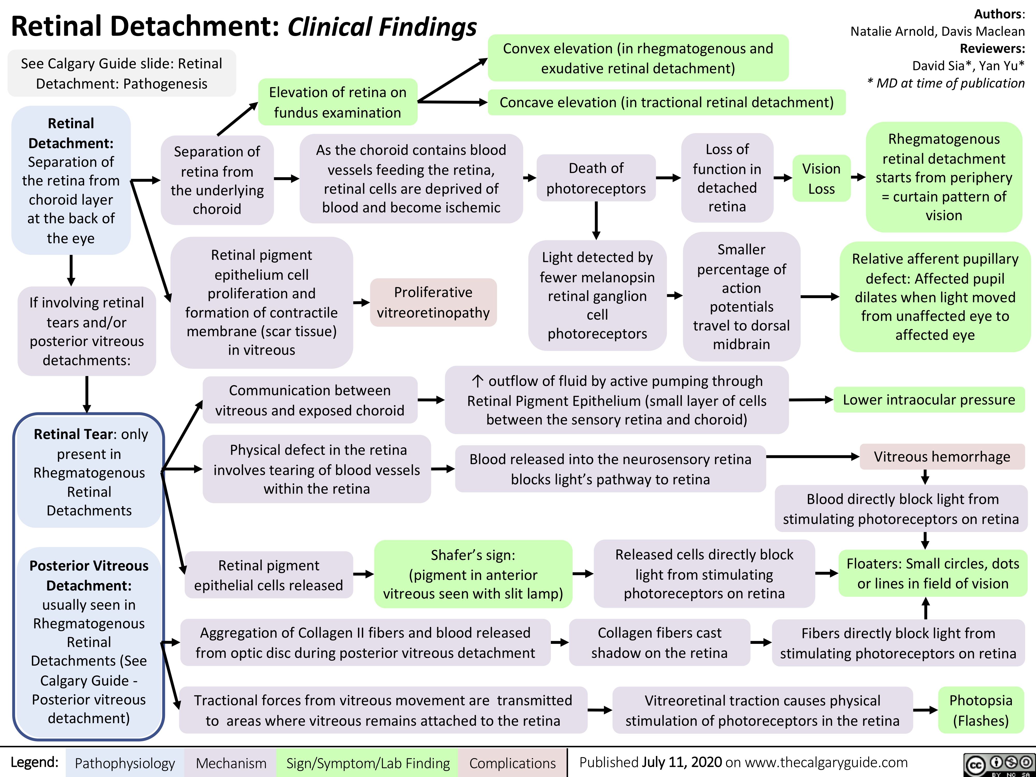
Retinal Detachment: Clinical Findings See Calgary Guide slide: Retinal
Convex elevation (in rhegmatogenous and exudative retinal detachment)
Concave elevation (in tractional retinal detachment)
Authors: Natalie Arnold, Davis Maclean Reviewers: David Sia*, Yan Yu* * MD at time of publication
Rhegmatogenous retinal detachment starts from periphery = curtain pattern of vision
Relative afferent pupillary defect: Affected pupil dilates when light moved from unaffected eye to affected eye
Lower intraocular pressure Vitreous hemorrhage
Detachment: Pathogenesis
Elevation of retina on fundus examination
Retinal Detachment: Separation of the retina from choroid layer at the back of the eye
If involving retinal tears and/or posterior vitreous detachments:
Retinal Tear: only present in Rhegmatogenous Retinal Detachments
Posterior Vitreous Detachment: usually seen in Rhegmatogenous Retinal Detachments (See Calgary Guide – Posterior vitreous detachment)
Separation of retina from the underlying choroid
As the choroid contains blood vessels feeding the retina, retinal cells are deprived of blood and become ischemic
Death of photoreceptors
Light detected by fewer melanopsin retinal ganglion cell photoreceptors
Loss of function in detached retina
Smaller percentage of action potentials travel to dorsal midbrain
Vision Loss
Retinal pigment epithelium cell proliferation and formation of contractile membrane (scar tissue) in vitreous
Proliferative vitreoretinopathy
Communication between vitreous and exposed choroid
Physical defect in the retina involves tearing of blood vessels within the retina
↑ outflow of fluid by active pumping through Retinal Pigment Epithelium (small layer of cells between the sensory retina and choroid)
Blood released into the neurosensory retina blocks light’s pathway to retina
Blood directly block light from stimulating photoreceptors on retina
Retinal pigment epithelial cells released
Shafer’s sign: (pigment in anterior vitreous seen with slit lamp)
Released cells directly block light from stimulating photoreceptors on retina
Floaters: Small circles, dots or lines in field of vision
Aggregation of Collagen II fibers and blood released from optic disc during posterior vitreous detachment
Tractional forces from vitreous movement are transmitted to areas where vitreous remains attached to the retina
Collagen fibers cast shadow on the retina
Fibers directly block light from stimulating photoreceptors on retina
Vitreoretinal traction causes physical Photopsia stimulation of photoreceptors in the retina (Flashes)
Legend:
Pathophysiology
Mechanism
Sign/Symptom/Lab Finding
Complications
Published July 11, 2020 on www.thecalgaryguide.com