Pyogenic Brain Abscess on MRI: Findings and Pathogenesis Common pathogens such as staphylococcus and streptococcus bacteria exist in the
outside environment or on the skin
Hematogenous Spread Direct Spread
Pathogen travels to brain by bloodstream Pathogen travels to brain by ears or sinus
Pathogen enters the brain parenchyma by crossing the blood brain barrier (BBB) transcellularly, paracellularly or by a Trojan Horse mechanism
Authors: Omer Mansoor, Aly Valji, Nameerah Wajahat Reviewers: Mao Ding, Reshma Sirajee, James Scott* *MD at time of publication
R
Transcellular
Pathogen invades BBB cell directly to enter
Paracellular
Pathogen goes between BBB cells by disrupting tight junctions
Trojan Horse
Pathogen bypasses BBB by hiding inside a macrophage cell
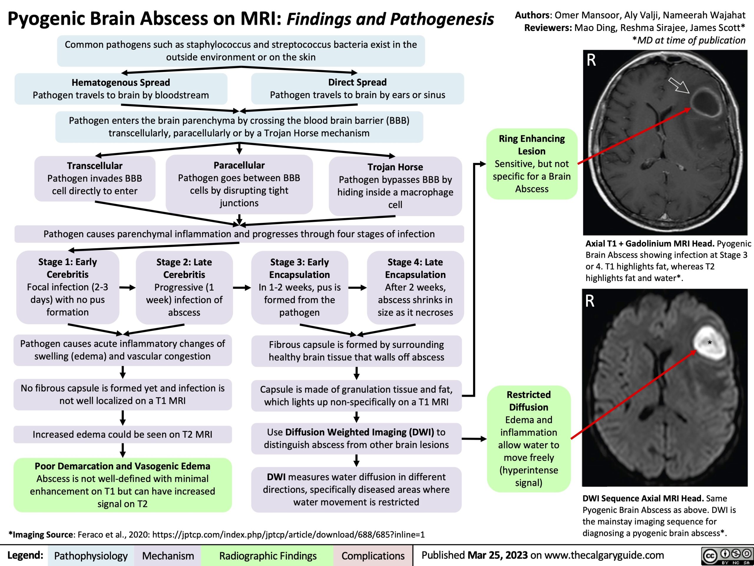
Ring Enhancing Lesion Sensitive, but not specific for a Brain Abscess
Pathogen causes parenchymal inflammation and progresses through four stages of infection
Axial T1 + Gadolinium MRI Head. Pyogenic Brain Abscess showing infection at Stage 3 or 4. T1 highlights fat, whereas T2 highlights fat and water*.
R
Stage 1: Early Cerebritis Focal infection (2-3 days) with no pus formation
Stage 2: Late Cerebritis Progressive (1 week) infection of abscess
Stage 3: Early Encapsulation
In 1-2 weeks, pus is formed from the pathogen
Stage 4: Late Encapsulation After 2 weeks, abscess shrinks in size as it necroses
Pathogen causes acute inflammatory changes of swelling (edema) and vascular congestion
No fibrous capsule is formed yet and infection is not well localized on a T1 MRI
Increased edema could be seen on T2 MRI
Poor Demarcation and Vasogenic Edema
Abscess is not well-defined with minimal enhancement on T1 but can have increased signal on T2
Fibrous capsule is formed by surrounding healthy brain tissue that walls off abscess
Capsule is made of granulation tissue and fat, which lights up non-specifically on a T1 MRI
Use Diffusion Weighted Imaging (DWI) to distinguish abscess from other brain lesions
DWI measures water diffusion in different directions, specifically diseased areas where water movement is restricted
Restricted Diffusion Edema and inflammation allow water to move freely (hyperintense signal)
*Imaging Source: Feraco et al., 2020: https://jptcp.com/index.php/jptcp/article/download/688/685?inline=1
DWI Sequence Axial MRI Head. Same Pyogenic Brain Abscess as above. DWI is the mainstay imaging sequence for diagnosing a pyogenic brain abscess*.
Legend:
Pathophysiology
Mechanism
Radiographic Findings
Complications
Published Mar 25, 2023 on www.thecalgaryguide.com