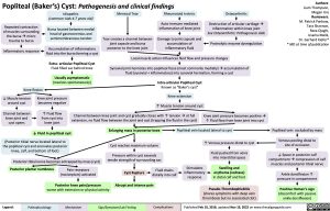
Popliteal (Baker’s) Cyst: Pathogenesis and clinical findings
Authors: Liam Thompson, Megan Ure Reviewers: M. Patrick Pankow, Tara Shannon, Reza Ojaghi, Usama Malik, Dr. Gerhard Kiefer* * MD at time of publication
Repeated contraction of muscles surrounding the bursaàmicro trauma to bursa
Inflammatory response
Idiopathic
(common: kids 4-7 years old)
Bursa located between medial head of gastrocnemius and semimembranosus tendon
Accumulation of inflammatory fluid into the bursa forming a cyst
Meniscal Tear
Tear creates a channel between joint capsule and bursa posterior to the knee joint
Rheumatoid Arthritis
Auto-immune mediated inflammation of knee joint
Osteoarthritis
Destruction of articular cartilageà inflammation within knee joint See Osteoarthritis: Pathogenesis slide
Damage to joint capsule and accumulation of inflammatory fluid
Proteolytic enzyme dysregulation Local muscle action influences fluid flow and pressure changes
Extra- articular Popliteal Cyst
Fluid filled sac behind knee
Usually asymptomatic
(resolves spontaneously)
Synovial joint herniates into popliteal fossa (most commonly medially)àaccumulation of fluid (synovial + inflammatory) into synovial herniation, forming a cyst
Knee flexion
Intra-articular Popliteal Cyst
Known as “Baker’s cyst”
Knee extension
↑ Muscle tension around cyst
Channel between knee joint and cyst gradually closes with ↑ tensionàat full extension, no fluid flow between the joint and cyst (trapping the fluid in the cyst)
↓ Muscle tension around cyst
Channel between knee joint and cyst opens
Knee joint pressure becomes negative
↑ Fluid flow from cyst into knee joint
↓ Fluid in popliteal cyst
Knee joint pressure becomes positiveà ↑ Fluid flow from knee joint into cyst
(Posterior tibial nerve located lateral to the popliteal cyst and enervates posterior knee, calf, and bottom of foot)
Posterior tibial nerve becomes entrapped by mass (cyst)
Enlarging mass in posterior knee
Cyst reaches maximum volume
Pressure within cyst exceeds tensile strength of surrounding sac
Fluid drains distally into calf
Popliteal vein located lateral to cyst
↑ Venous pressure distal to cyst
Popliteal vein occluded by mass
Venous pooling distal to site of occlusion
↓ Space in posterior calf compartmentàcompression of calf muscles and posterior tibial nerve
Ankle dorsiflexion ↑ pressure in compartment
Positive Homan’s sign
(discomfort with passive ankle dorsiflexion)
Posterior plantar numbness
Pain receptors (nociceptors) activated
Posterior knee pain/pressure
Cyst Rupture Abrupt and intense pain
Stimulates inflammatory response
Fluid pushed from veins into interstitial space
Swelling and erythema (redness) in distal calf and foot
Pseudo-Thrombophlebitis
worse with extension or physical activity
(shares symptoms with deep vein thrombosis but no associated clot)
Legend:
Pathophysiology
Mechanism
Sign/Symptom/Lab Finding
Complications
Published Feb 10, 2018, updated Nov 19, 2022 on www.thecalgaryguide.com
Foundations
Systems
Other Languages
Orthopedics Knee Popliteal (Baker’s) Cyst: Pathogenesis and clinical findings popliteal-bakers-cyst-pathogenesis-and-clinical-findings

