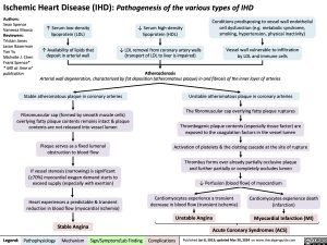
Ischemic Heart Disease (IHD): Pathogenesis of the various types of IHD
Authors:
Sean Spence Vaneeza Moosa Reviewers: Tristan Jones Jason Baserman Yan Yu
Michelle J. Chen Frank Spence* * MD at time of publication
↑ Serum low-density lipoprotein (LDL)
↑ Availability of lipids that deposit in arterial wall
↓ Serum high-density lipoprotein (HDL)
↓ LDL removal from coronary artery walls (transport of LDL to liver is impaired)
Atherosclerosis
Conditions predisposing to vessel wall endothelial cell dysfunction (e.g. metabolic syndrome, smoking, hypertension, physical inactivity)
Vessel wall vulnerable to infiltration by LDL and immune cells
Arterial wall degeneration, characterized by fat deposition (atheromatous plaque) in and fibrosis of the inner layer of arteries
Stable atheromatous plaque in coronary arteries
Fibromuscular cap (formed by smooth muscle cells) overlying fatty plaque contents remains intact & plaque contents are not released into vessel lumen
Plaque serves as a fixed lumenal obstruction to blood flow
If vessel stenosis (narrowing) is significant (≥70%) myocardial oxygen demand starts to exceed supply (especially with exertion)
Heart experiences a predictable & transient reduction in blood flow (myocardial ischemia)
Unstable atheromatous plaque in coronary arteries
The fibromuscular cap overlying fatty plaque ruptures
Thrombogenic plaque contents (especially tissue factor) are exposed to the coagulation factors in the vessel lumen
Activation of platelets & the clotting cascade at the site of rupture
Thrombus forms over already partially occlusive plaque and further partially or completely occludes lumen
↓ Perfusion (blood flow) of myocardium
Cardiomyocytes experience a transient decrease in blood flow (transient ischemia)
Unstable Angina
Cardiomyocytes experience death (infarction)
Myocardial Infarction (MI)
Stable Angina
Acute Coronary Syndromes (ACS)
Legend:
Pathophysiology
Mechanism
Sign/Symptom/Lab Finding
Complications
Published Jan 8, 2013; updated Mar 30, 2024 on www.thecalgaryguide.com
Foundations
Systems
Other Languages
Cardiology Chest Discomfort (Ischemic Heart Disease) Overview of Ischemic Heart Disease Overview of Ischemic Heart Disease

