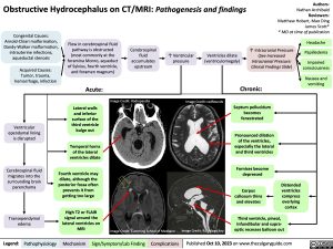
Obstructive Hydrocephalus on CT/MRI: Pathogenesis and findings
Authors: Nathan Archibald Reviewers: Matthew Hobart, Mao Ding James Scott* * MD at time of publication
Congenital Causes: Arnold-Chiari malformation, Dandy-Walker malformation, intrauterine infections, aqueductal stenosis
Acquired Causes: Tumor, trauma, hemorrhage, infection
Flow in cerebrospinal fluid pathway is obstructed (most commonly at the foramina Monro, aqueduct of Sylvius, fourth ventricle, and foramen magnum)
Acute:
Lateral walls and inferior surface of the third ventricle bulge out
Temporal horns Temporal horns
of the lateral of the lateral
ventricles dilate ventricles dilate
Cerebrospinal fluid accumulates upstream
↑ Ventricular pressure
Ventricles dilate (ventriculomegaly)
↑ Intracranial Pressure (See Increased Intracranial Pressure: Clinical Findings Slide)
Headache Papilledema
Impaired consciousness
Nausea and vomiting
Image Credit: Radiopaedia
Image Credit: radRounds
Ventricular ependymal lining is disrupted
Cerebrospinal fluid migrates into the surrounding brain parenchyma
Transependymal edema
Fourth ventricle may Fourth ventricle may
Chronic:
Septum pellucidum becomes fenestrated
Pronounced dilation of the ventricles, especially the lateral and third ventricles
Fornices become depressed
Corpus callosum thins and elevates
dilate, although the dilate, although the
posterior fossa often posterior fossa often
Distended ventricles compress overlying cortex
prevents it from prevents it from
getting too large getting too large
High T2 or FLAIR High T2 or FLAIR
signal around the signal around the
lateral ventricles on lateral ventricles on
MRI MRI
Image Credit: Cumming School of Medicine
Image Credit: Radiology Key
Third ventricle, pineal, infundibular and supra- optic recesses balloon out
Legend:
Pathophysiology
Mechanism
Sign/Symptom/Lab Finding
Complications
Published Oct 10, 2023 on www.thecalgaryguide.com
Foundations
Systems
Other Languages
Radiology Neuroradiology Obstructive Hydrocephalus on CT/MRI: Pathogenesis and findings Obstructive Hydrocephalus on CT MRI Pathogenesis and findings

