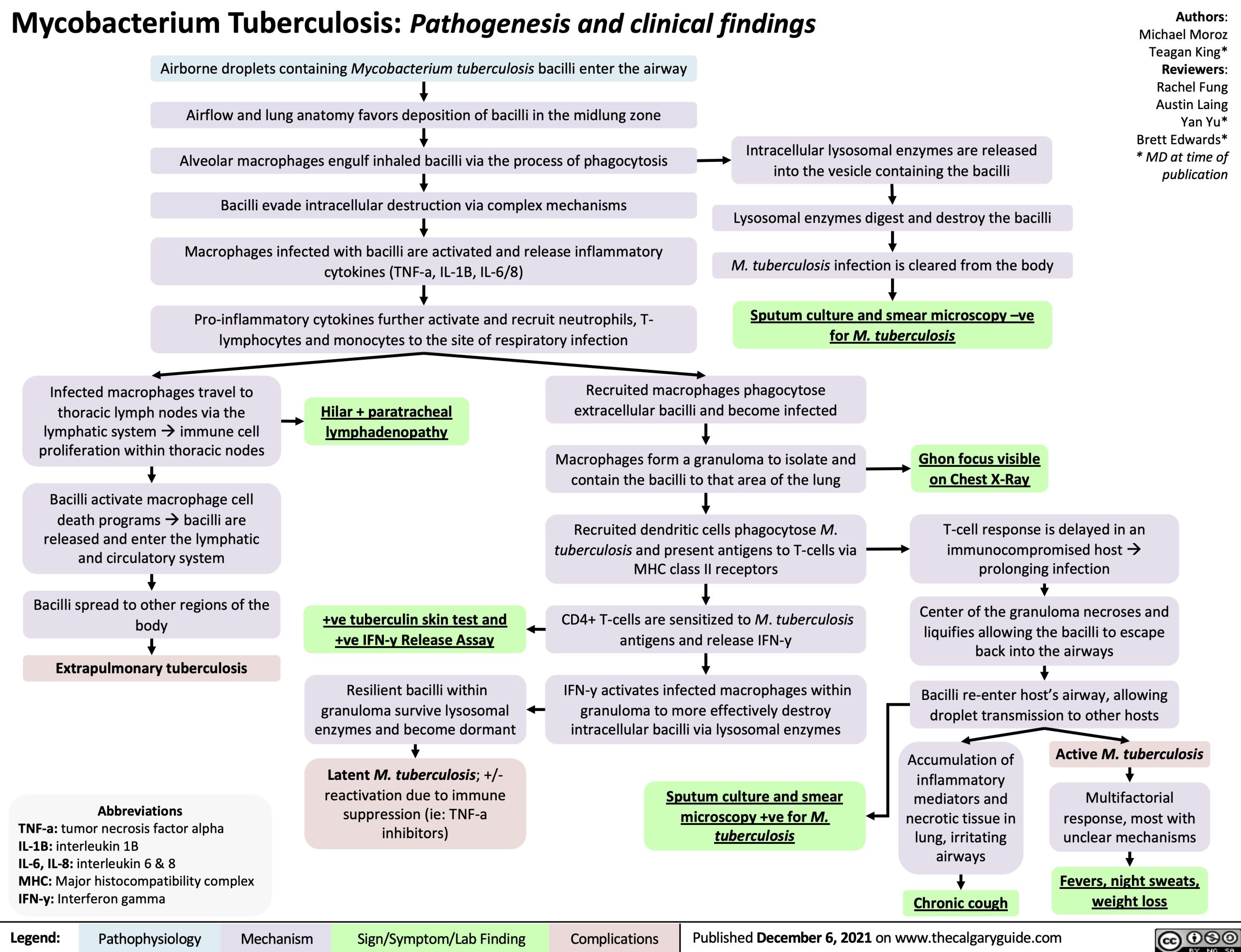
Mycobacterium Tuberculosis: Pathogenesis and clinical findings Airborne droplets containing Mycobacterium tuberculosis bacilli enter the airway
Authors: Michael Moroz Teagan King* Reviewers: Rachel Fung Austin Laing Yan Yu* Brett Edwards* * MD at time of publication
Airflow and lung anatomy favors deposition of bacilli in the midlung zone Alveolar macrophages engulf inhaled bacilli via the process of phagocytosis Bacilli evade intracellular destruction via complex mechanisms Macrophages infected with bacilli are activated and release inflammatory
cytokines (TNF-a, IL-1B, IL-6/8)
Pro-inflammatory cytokines further activate and recruit neutrophils, T- lymphocytes and monocytes to the site of respiratory infection
Intracellular lysosomal enzymes are released into the vesicle containing the bacilli
Lysosomal enzymes digest and destroy the bacilli M. tuberculosis infection is cleared from the body
Sputum culture and smear microscopy –ve for M. tuberculosis
Infected macrophages travel to thoracic lymph nodes via the lymphatic systemàimmune cell proliferation within thoracic nodes
Bacilli activate macrophage cell death programsàbacilli are
released and enter the lymphatic and circulatory system
Bacilli spread to other regions of the body
Extrapulmonary tuberculosis
Abbreviations TNF-a: tumor necrosis factor alpha
IL-1B: interleukin 1B
IL-6, IL-8: interleukin 6 & 8
MHC: Major histocompatibility complex IFN-y: Interferon gamma
Hilar + paratracheal lymphadenopathy
Recruited macrophages phagocytose extracellular bacilli and become infected
Macrophages form a granuloma to isolate and contain the bacilli to that area of the lung
Recruited dendritic cells phagocytose M. tuberculosis and present antigens to T-cells via MHC class II receptors
CD4+ T-cells are sensitized to M. tuberculosis antigens and release IFN-y
IFN-y activates infected macrophages within granuloma to more effectively destroy intracellular bacilli via lysosomal enzymes
Sputum culture and smear microscopy +ve for M. tuberculosis
Ghon focus visible on Chest X-Ray
+ve tuberculin skin test and +ve IFN-y Release Assay
Resilient bacilli within granuloma survive lysosomal enzymes and become dormant
Latent M. tuberculosis; +/- reactivation due to immune suppression (ie: TNF-a inhibitors)
T-cell response is delayed in an immunocompromised hostà prolonging infection
Center of the granuloma necroses and liquifies allowing the bacilli to escape back into the airways
Bacilli re-enter host’s airway, allowing droplet transmission to other hosts
Accumulation of inflammatory mediators and necrotic tissue in lung, irritating airways
Chronic cough
Active M. tuberculosis Multifactorial
response, most with unclear mechanisms
Fevers, night sweats, weight loss
Legend:
Pathophysiology
Mechanism
Sign/Symptom/Lab Finding
Complications
Published December 6, 2021 on www.thecalgaryguide.com