Classic Findings of Multiple Sclerosis (MS) on Brain MRI
Authors: Evan Allarie Davis Maclean Viesha Ciura* Reviewers: Yan Yu* * MD at time of publication
infiltrates in the infratentorial region
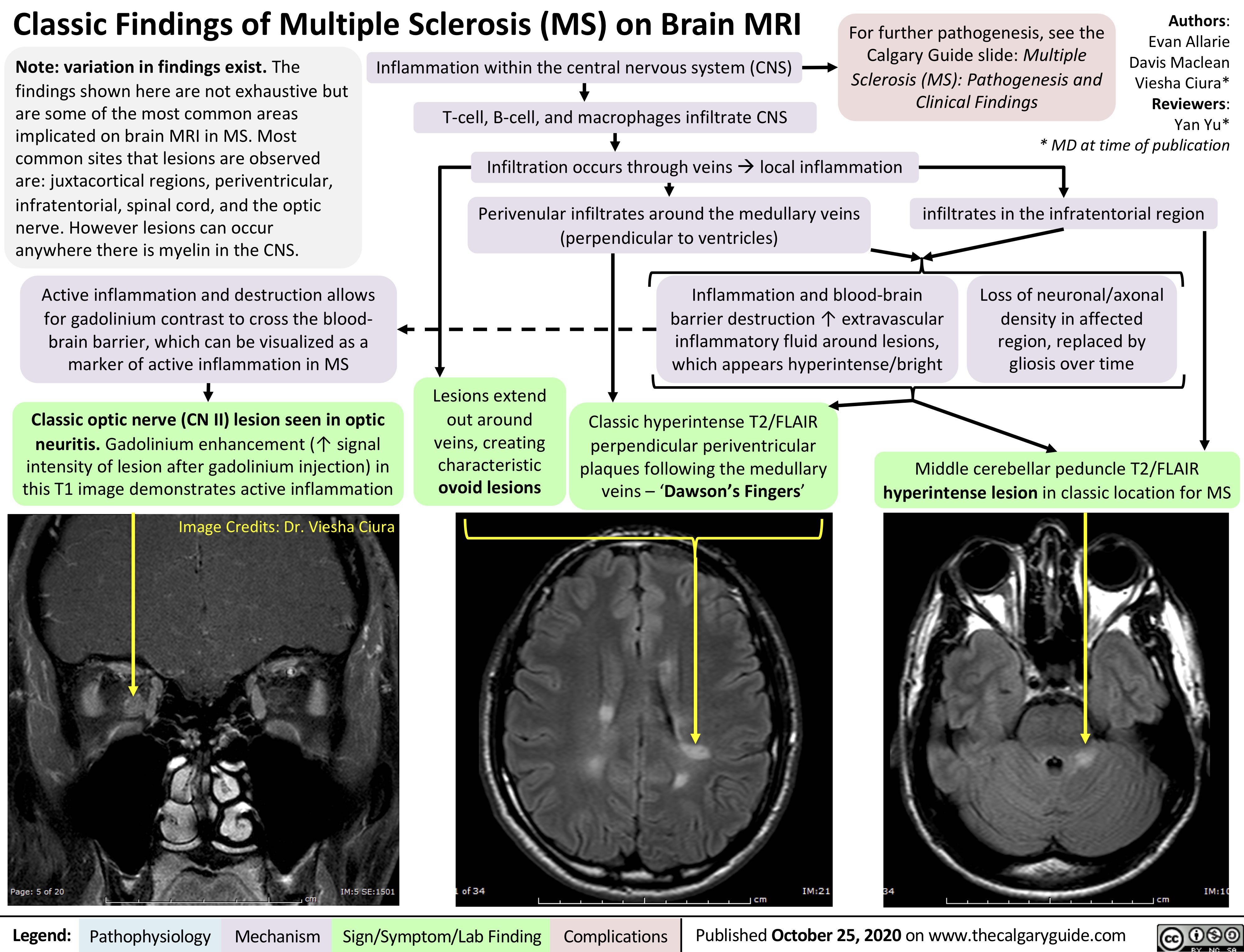
Note: variation in findings exist. The findings shown here are not exhaustive but are some of the most common areas implicated on brain MRI in MS. Most common sites that lesions are observed are: juxtacortical regions, periventricular, infratentorial, spinal cord, and the optic nerve. However lesions can occur anywhere there is myelin in the CNS.
Active inflammation and destruction allows for gadolinium contrast to cross the blood- brain barrier, which can be visualized as a marker of active inflammation in MS
Classic optic nerve (CN II) lesion seen in optic neuritis. Gadolinium enhancement (↑ signal
intensity of lesion after gadolinium injection) in this T1 image demonstrates active inflammation
Image Credits: Dr. Viesha Ciura
For further pathogenesis, see the Calgary Guide slide: Multiple Sclerosis (MS): Pathogenesis and Clinical Findings
Inflammation within the central nervous system (CNS) T-cell, B-cell, and macrophages infiltrate CNS
Infiltration occurs through veinsàlocal inflammation Perivenular infiltrates around the medullary veins
(perpendicular to ventricles)
Inflammation and blood-brain barrier destruction ↑ extravascular inflammatory fluid around lesions, which appears hyperintense/bright
Loss of neuronal/axonal density in affected region, replaced by gliosis over time
Lesions extend out around veins, creating characteristic ovoid lesions
Classic hyperintense T2/FLAIR perpendicular periventricular plaques following the medullary veins – ‘Dawson’s Fingers’
Middle cerebellar peduncle T2/FLAIR hyperintense lesion in classic location for MS
Legend:
Pathophysiology
Mechanism
Sign/Symptom/Lab Finding
Complications
Published October 25, 2020 on www.thecalgaryguide.com