Yu Yan – JVP explained – FINAL.pptx
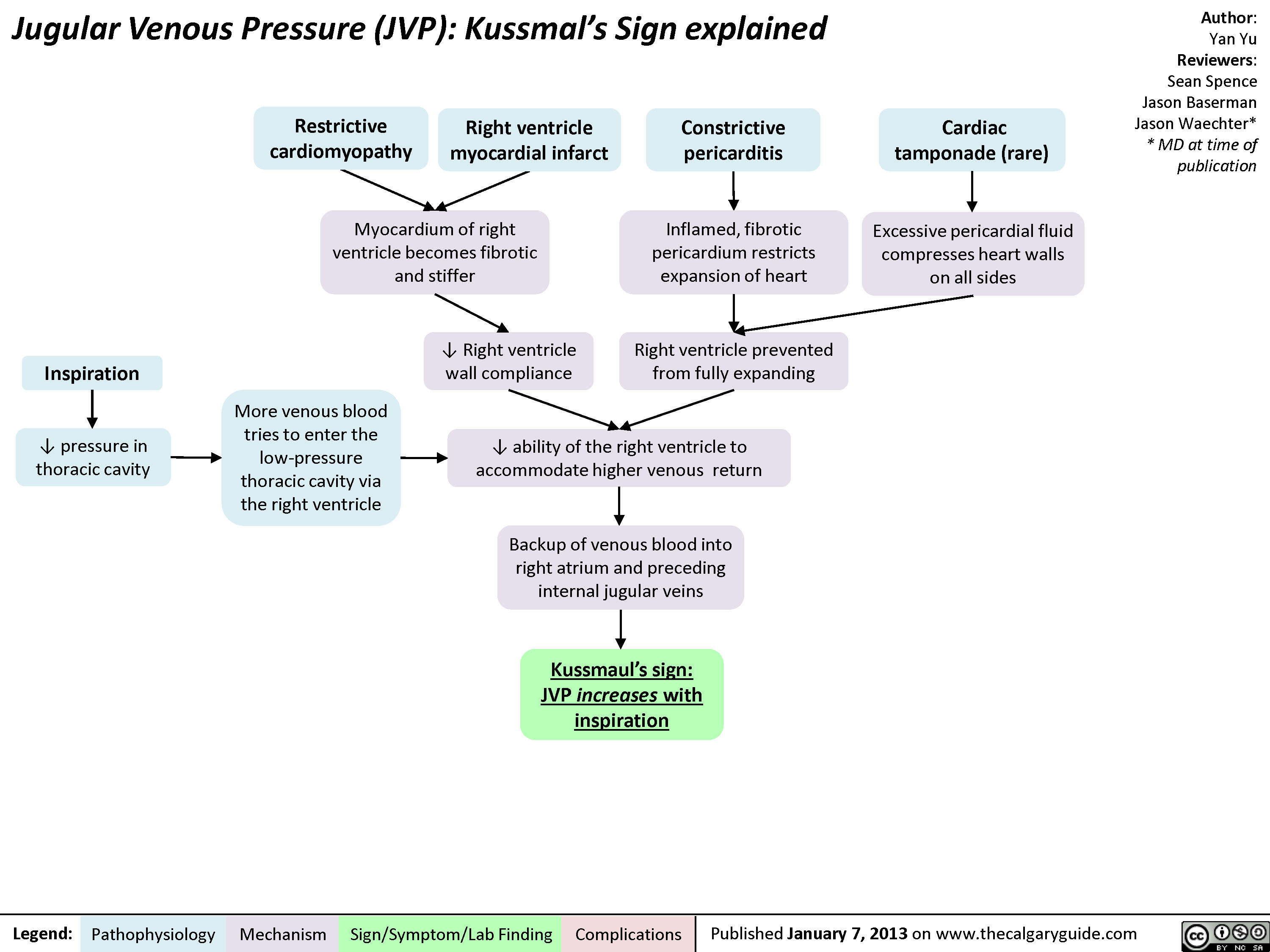
Myocardium of right ventricle becomes fibrotic and stifferKussmaul’s sign: JVP increases with inspirationJugular Venous Pressure (JVP): Kussmal’s Sign explainedExcessive pericardial fluid compresses heart walls on all sidesLegend:Published January 7, 2013 on www.thecalgaryguide.comMechanismPathophysiologySign/Symptom/Lab FindingComplicationsAuthor: Yan YuReviewers:Sean SpenceJason BasermanJason Waechter** MD at time of publication? Right ventricle wall complianceConstrictive pericarditisRight ventricle prevented from fully expanding ? ability of the right ventricle to accommodate higher venous returnBackup of venous blood into right atrium and preceding internal jugular veinsRestrictive cardiomyopathyInflamed, fibrotic pericardium restricts expansion of heartRight ventricle myocardial infarct Cardiac tamponade (rare)InspirationMore venous blood tries to enter the low-pressure thoracic cavity via the right ventricle? pressure in thoracic cavity
JVP should return to normal within 3 respiration cyclesJugular Venous Pressure (JVP): Physical Exam FeaturesExerts less force against vesselsTilting the head of the bed:Low pressure, & the thinner walls of the internal jugular veins, are less able to keep lumen open when compressedLegend:Published January 7, 2013 on www.thecalgaryguide.comMechanismPathophysiologySign/Symptom/Lab FindingComplicationsAuthor: Yan YuReviewers:Sean SpenceJason BasermanJason Waechter** MD at time of publicationBlood in internal jugulars settles to bottom of the vein (analogy: half-full tube of water is turned vertically)Visible waves in the JVP correspond to stages of the cardiac cycleNon-palpableBiphasic waveform? JVP The Jugular Venous Pressure (JVP) Blood pressure in the internal jugular veinsV-waveA-waveRight atrial contractionBlood passively fills right atrium during ventricular systole? JVP? JVPIn: ? intrathoracic pressurePressing hard on abdomen (overlying the liver), or doing a valsalvaFacing lower afterload, Right heart more readily pumps blood into pulmonary circulation? abdominal pressure? venous blood forced up into right atriumVenous blood pressure is normally very lowOccludableNote: Since the internal jugular veins are continuous with the right atrium, the JVP is a reliable estimate of right atrial blood pressure (Central Venous Pressure). The JVP on the right side is a better “barometer” than the JVP on the left, because the R internal jugular communicates more directly with the R atrium.Note: The Jugular Venous Pressure is also called the “jugular venous pulse”. ? filling pressure of right atrium and internal jugular veinsJVP changes with body positionUpBlood in internal jugulars settles along the side of the vein (analogy: half-full tube of water is turned horizontally)? JVP DownJVP changes with respiration? JVPOut: ? intrathoracic pressureFacing higher afterload, Right heart is less able to pump blood into pulmonary circulation? filling pressure of right atrium and internal jugular veinsBreathing:? blood back-up into internal jugular veinsPulmonary circulation accepts more venous blood (especially on inspiration, when the thoracic cavity expands)Heart fills with more blood (? preload), ? stroke volume of next heart-beat, shifting more blood into arteries? pressure in right atrium is transmitted into internal jugular veins, ?JVP
120 kB / 440 words