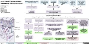
Deep Partial Thickness Burns:
Pathogenesis and clinical findings
Radiation (Sunlight, x-ray, nuclear emission/explosion)
Ionizing radiation gets into contact with DNA
Damage to keratinocytes
Fire (Flash fire or direct contact with flame)
Contact (Hot solid objects)
Scalding (Hot liquid via immersion, spill or splash)
Chemical (Strong acid, alkali or irritant gas)
Electrical (Contact with exposed electrical wiring/appliances)
Author and Illustrator: Amanda Eslinger Maharshi Gandhi Reviewers:
Alexander Arnold
Shahab Marzoughi
Duncan Nickerson*
* MD at time of publication
Direct transfer of heat energy
Coagulation necrosis (cell death due to ischemia) is induced
Deep Partial Thickness Burn
Injury to the epidermal layer and both the papillary and a portion of the reticular layer of the dermis
Transfer of heat energy & direct injury to cellular membranes
Epidermis
Papillary dermis Reticular dermis
Sub- cutaneous Tissue
↑ vascular permeability due to damage
Fluid leak results in edema between dermal & epidermal layer
Blisters (a bubble on the skin containing fluid)
Thin epidermal layer forming fluid-filled vesicle breaks open
Cutaneous capillary bed is destroyed
Blood flow to injured area is compromised
Pressing on the skin doesn’t easily force blood cells out of the area
Non-blanchable skin
Vasodilation in fascia (a thin casing of connective tissue) underlying subcutaneous tissue
Inflammatory mediators such as cytokines and prostaglandins activate fibroblasts
Collagen deposition and subsequently induration (thickening of skin)
Some healthy dermal appendages surrounded by islands of undamaged epithelial cells
Possible spontaneous re-epithelialization in 2-9 weeks
During re-epithelization of burns there can be excessive deposition of collagen and other extracellular matrix components
Thick raised scars in dermis due to excess collagen deposition
Somatosensory structures (nociceptors, thermoreceptors, and mechanoreceptors) are completely injured within the dermis
Analgesia of impacted area (the inability to feel pain)
Ongoing Inflammation and dilated blood vessels
Red Induration (Thickened skin appearing red)
Compromised blood flow and more extensive tissue damage
White Induration (Thickened skin appearing white)
Moist Wound
Legend:
Pathophysiology
Mechanism
Sign/Symptom/Lab Finding
Complications
Published Dec 2, 2013, updated Apr 20, 2024 on www.thecalgaryguide.com
Foundations
Systems
Other Languages
Dermatology Burns Deep Partial Thickness Burns: Pathogenesis and Clinical Findings Deep Partial Thickness Burns Pathogenesis and Clinical Findings

