Circle of Willis: Anatomy and Physiology
Authors: Josh Kariath Reviewers: Andrea Kuczynski Gary Klein* * MD at time of publication
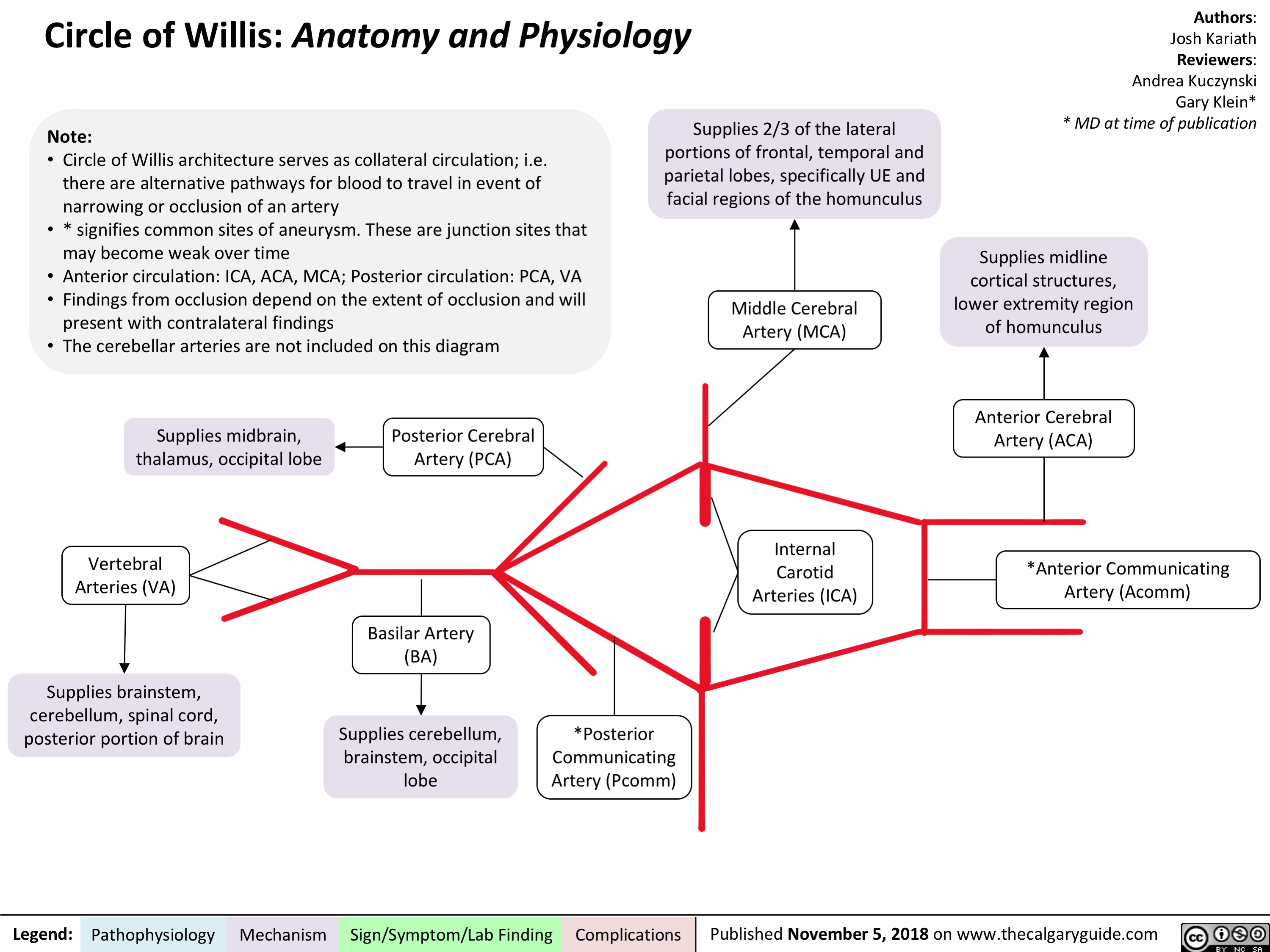
Supplies midline cortical structures, lower extremity region of homunculus
Anterior Cerebral Artery (ACA)
*Anterior Communicating Artery (Acomm)
Note:
• Circle of Willis architecture serves as collateral circulation; i.e. there are alternative pathways for blood to travel in event of narrowing or occlusion of an artery
• * signifies common sites of aneurysm. These are junction sites that may become weak over time
• Anterior circulation: ICA, ACA, MCA; Posterior circulation: PCA, VA
• Findings from occlusion depend on the extent of occlusion and will
present with contralateral findings
• The cerebellar arteries are not included on this diagram
Supplies 2/3 of the lateral portions of frontal, temporal and parietal lobes, specifically UE and facial regions of the homunculus
Middle Cerebral Artery (MCA)
Supplies midbrain, thalamus, occipital lobe
Vertebral Arteries (VA)
Supplies brainstem, cerebellum, spinal cord, posterior portion of brain
Posterior Cerebral Artery (PCA)
Legend: Pathophysiology Mechanism
Sign/Symptom/Lab Finding Complications Published November 5, 2018 on www.thecalgaryguide.com
Basilar Artery (BA)
Supplies cerebellum, brainstem, occipital lobe
*Posterior Communicating Artery (Pcomm)
Internal Carotid Arteries (ICA)