Avascular Necrosis (AVN) of the Femoral Head:
Findings on X-Ray
Blood supply to the subchondral bone is disrupted (for full pathogenesis, see Calgary Guide slide Avascular Necrosis: Pathogenesis and clinical findings)
Hypoxia at boneàOsteocyte (primary bone cell) apoptosis
Authors: Daniel Cusano, Evan Allarie, Reviewers: Yan Yu* Davis Maclean, Shelley Spaner* *MD at time of publication
↓ local osteoprotegerin (OPG) production
OPG acts to 1) inhibit osteoclast formation, & 2) bind and sequester RANKL, a chemical that stimulates osteoclast activation & proliferation
↓ OPGà↑ osteoclast activation & proliferationà↑ bone resorption
Areas that absorb ↑ X-ray radiation appear brighter on X-ray
RANKL – Receptor activator of nuclear factor kappa-Β ligand
Basic X-Ray Physiology
Bone absorbs more X-ray radiation than other tissues in the body (e.g fat and muscle)
Areas of ↓ bone density are darker: described as (radio)lucency on X-ray
Areas of ↑ bone density are whiter: described as sclerotic on X-ray
Image Credit: Alberta Health Services Repository
↓ Production of vascular endothelial growth factor (VEGF), which normally maintains & builds healthy bone & vasculature (via a multifactorial process)
Osteopenia: generalized ↓ in bone density, visualized as ↑ radiolucency (darkness) of bone
Bone demineralization and collagen matrix destruction
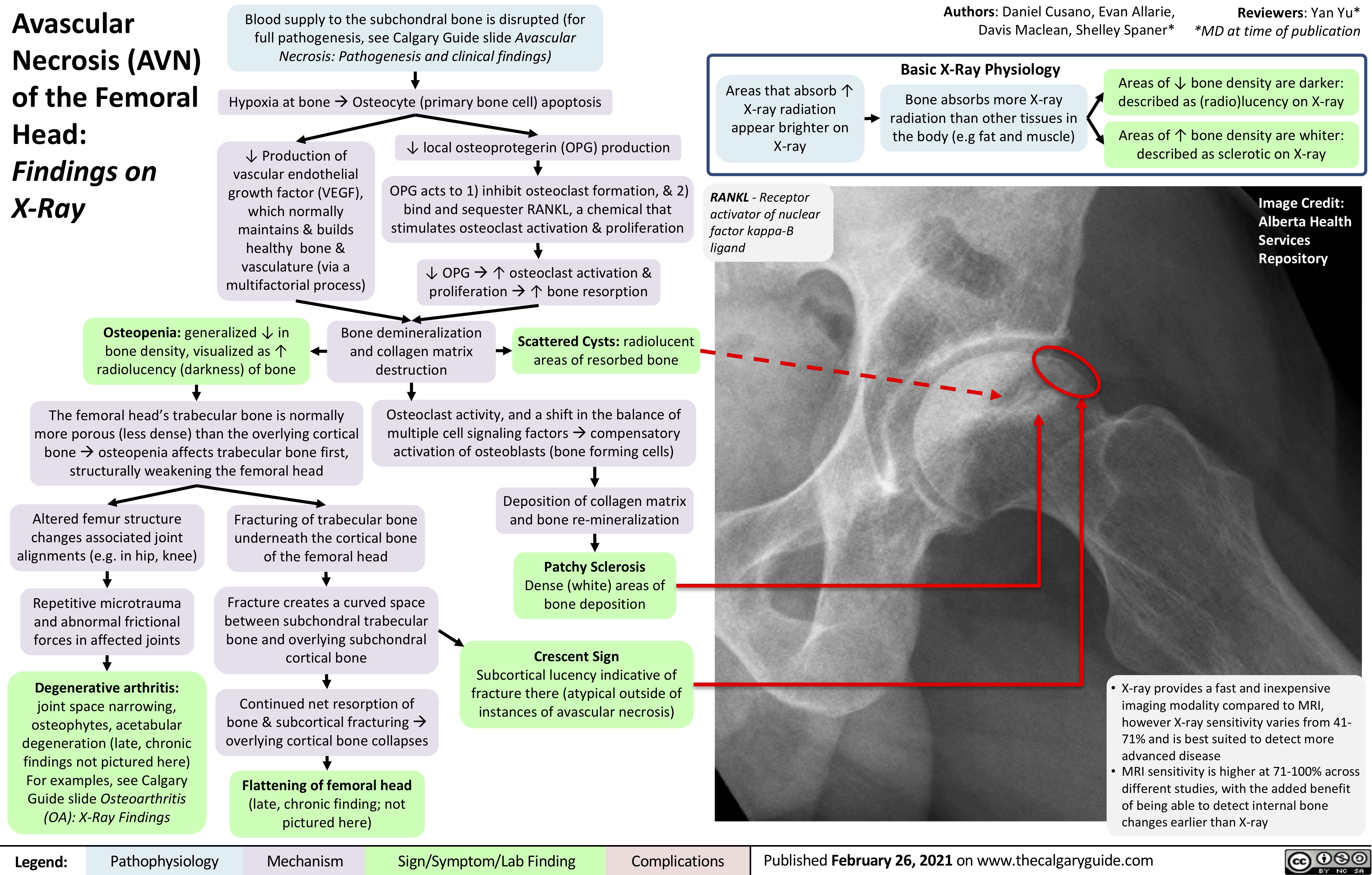
Scattered Cysts: radiolucent areas of resorbed bone
The femoral head’s trabecular bone is normally more porous (less dense) than the overlying cortical boneàosteopenia affects trabecular bone first, structurally weakening the femoral head
Osteoclast activity, and a shift in the balance of multiple cell signaling factorsàcompensatory activation of osteoblasts (bone forming cells)
Altered femur structure changes associated joint alignments (e.g. in hip, knee)
Repetitive microtrauma and abnormal frictional forces in affected joints
Degenerative arthritis:
joint space narrowing, osteophytes, acetabular degeneration (late, chronic findings not pictured here) For examples, see Calgary Guide slide Osteoarthritis (OA): X-Ray Findings
Fracturing of trabecular bone underneath the cortical bone of the femoral head
Fracture creates a curved space between subchondral trabecular bone and overlying subchondral cortical bone
Continued net resorption of bone & subcortical fracturingà overlying cortical bone collapses
Flattening of femoral head
(late, chronic finding; not pictured here)
Deposition of collagen matrix and bone re-mineralization
Patchy Sclerosis
Dense (white) areas of bone deposition
Crescent Sign
Subcortical lucency indicative of fracture there (atypical outside of instances of avascular necrosis)
• X-ray provides a fast and inexpensive imaging modality compared to MRI, however X-ray sensitivity varies from 41- 71% and is best suited to detect more advanced disease
• MRI sensitivity is higher at 71-100% across different studies, with the added benefit of being able to detect internal bone changes earlier than X-ray
Legend:
Pathophysiology
Mechanism
Sign/Symptom/Lab Finding
Complications
Published February 26, 2021 on www.thecalgaryguide.com