Yu Yan – AFib Clinical Findings – FINAL.pptx
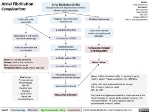
Atrial Fibrillation: Clinical FindingsAbnormal electrical signals in fibrillating atria can propagate to ventricles before ventricles have fully recovered from their previous contraction.Uncoordinated, irregular atrial contraction? Diastolic ventricular filling timeLegend:Published January 22. 2013 on www.thecalgaryguide.comMechanismPathophysiologySign/Symptom/Lab FindingComplicationsAuthor: Yan YuReviewers:Rob SchultzSean SpenceNanette Alvarez** MD at time of publicationNo discrete “Atrial kick” at the end of ventricular diastoleMore forceful contractions of sometimes overfilled ventricles are felt against the chest wall Preload ?? cardiac outputPalpitationsDyspnea (shortness of breath)No S4 on auscultation(even with non-compliant ventricle)Atrial fibrillation (A-fib)Disorganized atrial depolarizations without effective atrial contraction.Presyncopeor SyncopeFatigue? Perfusion of brain and other body tissuesHypoxia causes respiratory centers in brain to compensate by ? rate of breathingPhysical exam:History:Disorganized atrial electrical circuitsIrregularly Irregular Pulse (often with ? heart rate)Even with non-compliant ventricles, atrial systole ejects minimal blood into ventricle with each contractionBlowing Holosystolic murmurVariable S1 loudnessAfter abnormal atrial contraction, AV valve leaflets can range from fully open to almost closedAV valve annular dilationIrregular, often faster relay of electrical impulses through AV node and purkinje fibersIrregular, often faster stimulation of ventricular contractionsSubsequent ven-tricular contractions force the AV valve leaflets shut at varying velocities, producing varying levels of soundMitral/tricuspid valve leaflets do not properly coaptBlood regurgitates through AV valves during ventricular contractionNext ventricular contraction is weaker; ? blood ejected (? ejection fraction)More residual blood in ventricles as diastole starts, ? end-diastolic volume prior to next ventricular contractionIf heart rate is increased:? time to clear Ca2+ from myocytes’ cytosol between contractions? in cytosolic [Ca2+] prior to subsequent myocyte contractions? force of contractionNote: Most patients with A-Fib are asymptomatic!L lower sternal border = Tricuspid regurgitation)Apex = Mitral regur-gitationNo “A-waves” in JVP
115 kB / 270 words

