Arachnoid Cysts: Pathogenesis and clinical findings
Authors: George S. Tadros Reviewers: Yvette Ysabel Yao Shahab Marzoughi Gary Michael Klein* * MD at time of publication
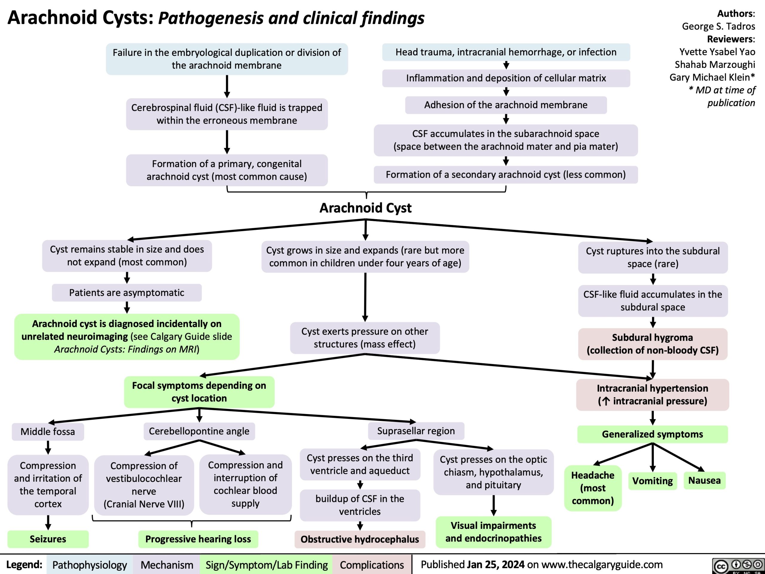
Failure in the embryological duplication or division of the arachnoid membrane
Cerebrospinal fluid (CSF)-like fluid is trapped within the erroneous membrane
Formation of a primary, congenital arachnoid cyst (most common cause)
Head trauma, intracranial hemorrhage, or infection
Inflammation and deposition of cellular matrix Adhesion of the arachnoid membrane
CSF accumulates in the subarachnoid space (space between the arachnoid mater and pia mater)
Formation of a secondary arachnoid cyst (less common)
Cyst remains stable in size and does not expand (most common)
Patients are asymptomatic
Arachnoid cyst is diagnosed incidentally on unrelated neuroimaging (see Calgary Guide slide Arachnoid Cysts: Findings on MRI)
Arachnoid Cyst
Cyst grows in size and expands (rare but more common in children under four years of age)
Cyst exerts pressure on other structures (mass effect)
Suprasellar region
Cyst ruptures into the subdural space (rare)
CSF-like fluid accumulates in the subdural space
Subdural hygroma (collection of non-bloody CSF)
Intracranial hypertension (↑ intracranial pressure)
Generalized symptoms
Middle fossa
Compression and irritation of the temporal cortex
Seizures
Focal symptoms depending on cyst location
Cerebellopontine angle
Compression of vestibulocochlear nerve (Cranial Nerve VIII)
Compression and interruption of cochlear blood supply
Cyst presses on the third ventricle and aqueduct
buildup of CSF in the ventricles
Obstructive hydrocephalus
Cyst presses on the optic chiasm, hypothalamus, and pituitary
Visual impairments and endocrinopathies
Headache (most common)
Vomiting
Nausea
Progressive hearing loss
Legend:
Pathophysiology
Mechanism
Sign/Symptom/Lab Finding
Complications
Published Jan 25, 2024 on www.thecalgaryguide.com