Arachnoid Cysts: Findings on MRI
Imaging source:
radiopaedia.org
Authors: George S. Tadros Reviewers: Matthew Hobart, Shahab Marzoughi, James Scott* * MD at time of publication
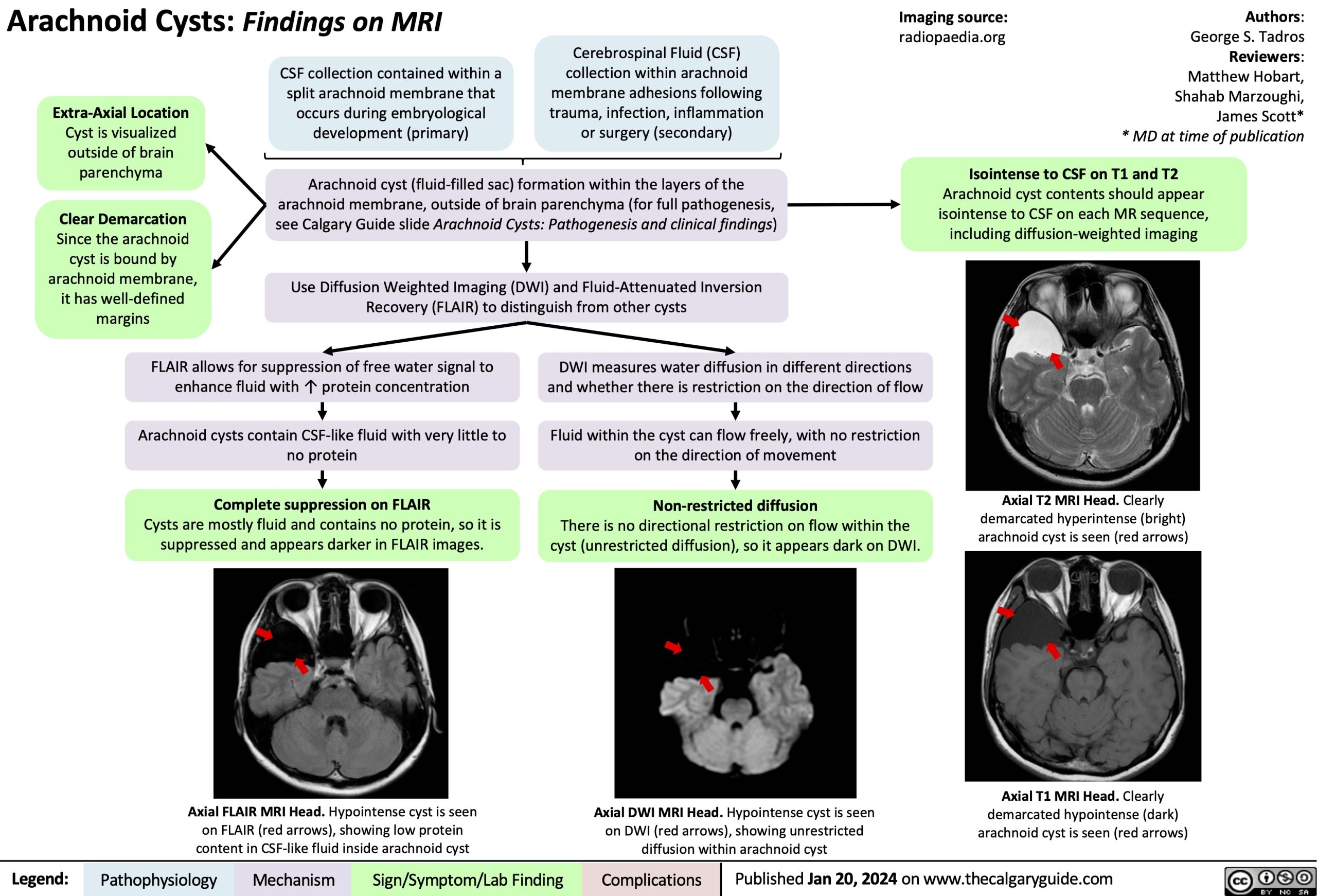
Extra-Axial Location
Cyst is visualized outside of brain parenchyma
Clear Demarcation
Since the arachnoid cyst is bound by arachnoid membrane, it has well-defined margins
CSF collection contained within a split arachnoid membrane that occurs during embryological development (primary)
Cerebrospinal Fluid (CSF) collection within arachnoid membrane adhesions following trauma, infection, inflammation or surgery (secondary)
Arachnoid cyst (fluid-filled sac) formation within the layers of the arachnoid membrane, outside of brain parenchyma (for full pathogenesis, see Calgary Guide slide Arachnoid Cysts: Pathogenesis and clinical findings)
Use Diffusion Weighted Imaging (DWI) and Fluid-Attenuated Inversion Recovery (FLAIR) to distinguish from other cysts
Isointense to CSF on T1 and T2
Arachnoid cyst contents should appear isointense to CSF on each MR sequence, including diffusion-weighted imaging
Axial T2 MRI Head. Clearly demarcated hyperintense (bright) arachnoid cyst is seen (red arrows)
Axial T1 MRI Head. Clearly demarcated hypointense (dark) arachnoid cyst is seen (red arrows)
FLAIR allows for suppression of free water signal to enhance fluid with ↑ protein concentration
Arachnoid cysts contain CSF-like fluid with very little to no protein
Complete suppression on FLAIR
Cysts are mostly fluid and contains no protein, so it is suppressed and appears darker in FLAIR images.
Axial FLAIR MRI Head. Hypointense cyst is seen on FLAIR (red arrows), showing low protein content in CSF-like fluid inside arachnoid cyst
DWI measures water diffusion in different directions and whether there is restriction on the direction of flow
Fluid within the cyst can flow freely, with no restriction on the direction of movement
Non-restricted diffusion
There is no directional restriction on flow within the cyst (unrestricted diffusion), so it appears dark on DWI.
Axial DWI MRI Head. Hypointense cyst is seen on DWI (red arrows), showing unrestricted diffusion within arachnoid cyst
Legend:
Pathophysiology
Mechanism
Sign/Symptom/Lab Finding
Complications
Published Jan 20, 2024 on www.thecalgaryguide.com