Alcoholic Fatty Liver Disease: Pathogenesis and clinical findings
Authors: Tara Shannon Reviewers: Ben Campbell, Yan Yu*, Samuel Lee* * MD at time of publication
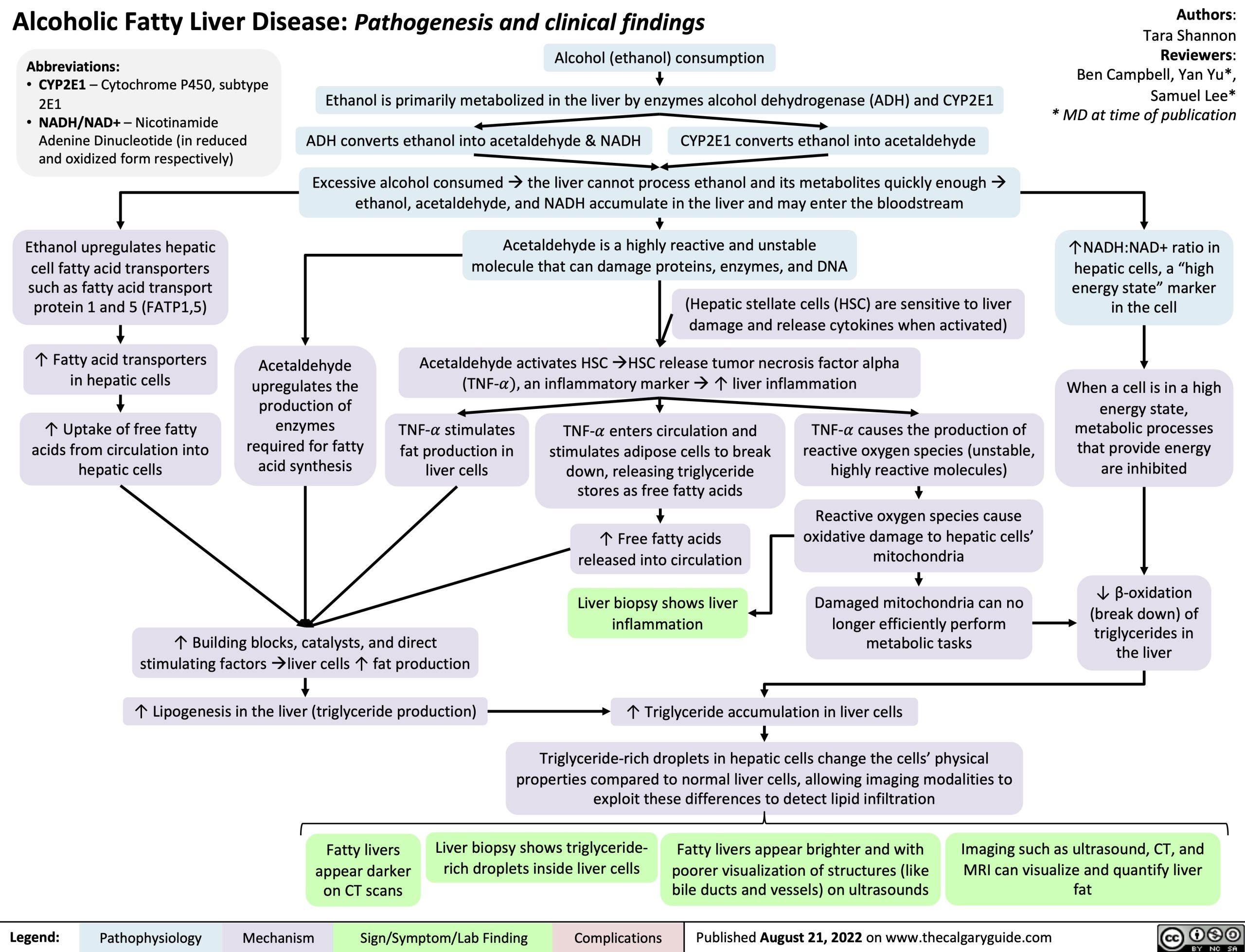
↑NADH:NAD+ ratio in hepatic cells, a “high energy state” marker in the cell
When a cell is in a high energy state, metabolic processes that provide energy are inhibited
↓ β-oxidation (break down) of
triglycerides in the liver
Abbreviations:
• CYP2E1 – Cytochrome P450, subtype
2E1
• NADH/NAD+ – Nicotinamide
Adenine Dinucleotide (in reduced and oxidized form respectively)
Ethanol upregulates hepatic cell fatty acid transporters
such as fatty acid transport protein 1 and 5 (FATP1,5)
↑ Fatty acid transporters in hepatic cells
↑ Uptake of free fatty acids from circulation into hepatic cells
Alcohol (ethanol) consumption
Ethanol is primarily metabolized in the liver by enzymes alcohol dehydrogenase (ADH) and CYP2E1 ADH converts ethanol into acetaldehyde & NADH CYP2E1 converts ethanol into acetaldehyde
Excessive alcohol consumedàthe liver cannot process ethanol and its metabolites quickly enoughà ethanol, acetaldehyde, and NADH accumulate in the liver and may enter the bloodstream
Acetaldehyde is a highly reactive and unstable molecule that can damage proteins, enzymes, and DNA
(Hepatic stellate cells (HSC) are sensitive to liver damage and release cytokines when activated)
Acetaldehyde activates HSCàHSC release tumor necrosis factor alpha (TNF-!), an inflammatory markerà↑ liver inflammation
Acetaldehyde upregulates the production of enzymes required for fatty acid synthesis
TNF-! stimulates fat production in liver cells
TNF-! enters circulation and stimulates adipose cells to break down, releasing triglyceride stores as free fatty acids
↑ Free fatty acids released into circulation
Liver biopsy shows liver inflammation
TNF-! causes the production of reactive oxygen species (unstable, highly reactive molecules)
Reactive oxygen species cause oxidative damage to hepatic cells’ mitochondria
Damaged mitochondria can no longer efficiently perform metabolic tasks
↑ Building blocks, catalysts, and direct stimulating factorsàliver cells ↑ fat production
↑ Lipogenesis in the liver (triglyceride production)
↑ Triglyceride accumulation in liver cells
Triglyceride-rich droplets in hepatic cells change the cells’ physical properties compared to normal liver cells, allowing imaging modalities to exploit these differences to detect lipid infiltration
Fatty livers appear darker on CT scans
Liver biopsy shows triglyceride- rich droplets inside liver cells
Fatty livers appear brighter and with poorer visualization of structures (like bile ducts and vessels) on ultrasounds
Imaging such as ultrasound, CT, and MRI can visualize and quantify liver fat
Legend:
Pathophysiology
Mechanism
Sign/Symptom/Lab Finding
Complications
Published August 21, 2022 on www.thecalgaryguide.com