Acute MCA-Territory Ischemic Stroke: Findings on Non-Contrast CT
See Calgary Guide slide – Ischemic Stroke: Pathogenesis
Middle cerebral artery (MCA) strokes are generally caused by emboli (blood clot that travelled from elsewhere in the body) and less commonly caused by thrombus (blood clot that has developed locally in the MCA)
Embolism (or thrombus) occludes flow through the MCA
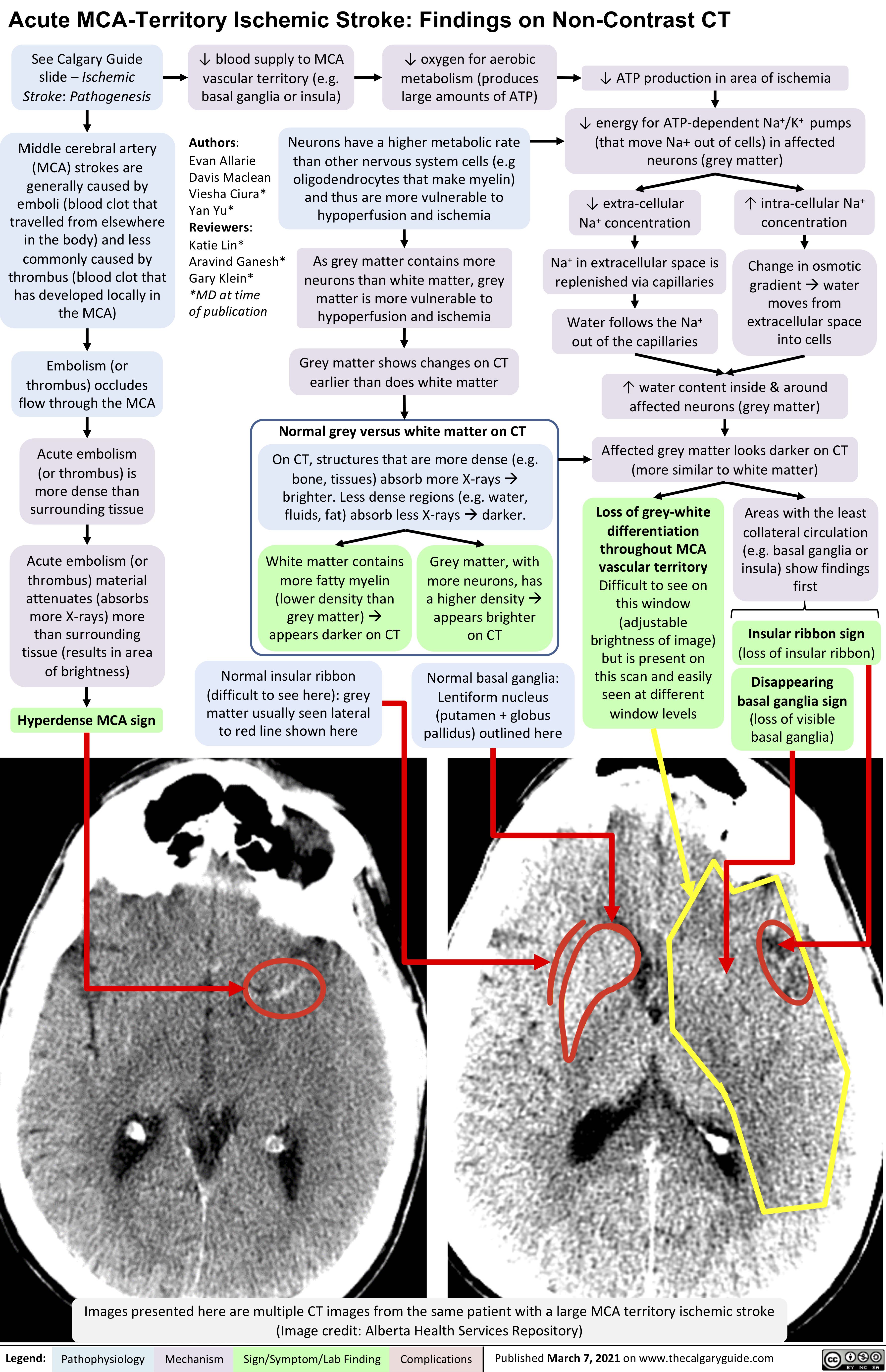
Acute embolism
(or thrombus) is more dense than surrounding tissue
Acute embolism (or thrombus) material attenuates (absorbs more X-rays) more than surrounding tissue (results in area of brightness)
Hyperdense MCA sign
↓ blood supply to MCA vascular territory (e.g. basal ganglia or insula)
↓ oxygen for aerobic metabolism (produces large amounts of ATP)
↓ ATP production in area of ischemia
↓ energy for ATP-dependent Na+/K+ pumps (that move Na+ out of cells) in affected neurons (grey matter)
Neurons have a higher metabolic rate than other nervous system cells (e.g oligodendrocytes that make myelin) and thus are more vulnerable to hypoperfusion and ischemia
As grey matter contains more neurons than white matter, grey matter is more vulnerable to hypoperfusion and ischemia
Grey matter shows changes on CT earlier than does white matter
Normal grey versus white matter on CT
On CT, structures that are more dense (e.g. bone, tissues) absorb more X-raysà brighter. Less dense regions (e.g. water, fluids, fat) absorb less X-raysàdarker.
Authors:
Evan Allarie Davis Maclean Viesha Ciura* Yan Yu* Reviewers:
Katie Lin* Aravind Ganesh* Gary Klein*
*MD at time
of publication
↓ extra-cellular Na+ concentration
Na+ in extracellular space is replenished via capillaries
Water follows the Na+ out of the capillaries
↑ intra-cellular Na+ concentration
Change in osmotic gradientàwater moves from extracellular space into cells
↑ water content inside & around affected neurons (grey matter)
Affected grey matter looks darker on CT (more similar to white matter)
White matter contains more fatty myelin (lower density than grey matter)à appears darker on CT
Normal insular ribbon (difficult to see here): grey matter usually seen lateral to red line shown here
Grey matter, with more neurons, has a higher densityà appears brighter on CT
Normal basal ganglia: Lentiform nucleus (putamen + globus pallidus) outlined here
Loss of grey-white differentiation throughout MCA vascular territory Difficult to see on this window (adjustable brightness of image) but is present on this scan and easily seen at different window levels
Areas with the least collateral circulation
(e.g. basal ganglia or insula) show findings first
Insular ribbon sign
(loss of insular ribbon)
Disappearing basal ganglia sign (loss of visible basal ganglia)
Comparison to normal anatomy helps outline pathologic findings
Comparison to normal anatomy helps outline pathologic findings
Images presented here are multiple CT images from the same patient with a large MCA territory ischemic stroke (Image credit: Alberta Health Services Repository)
Legend:
Pathophysiology
Mechanism
Sign/Symptom/Lab Finding
Complications
Published March 7, 2021 on www.thecalgaryguide.com