• Content
  • Collaboration
  • About Us
  • Contact Us

SEARCH RESULTS FOR: Sinusitis

Bronchiectasis Pathogenesis and clinical findings
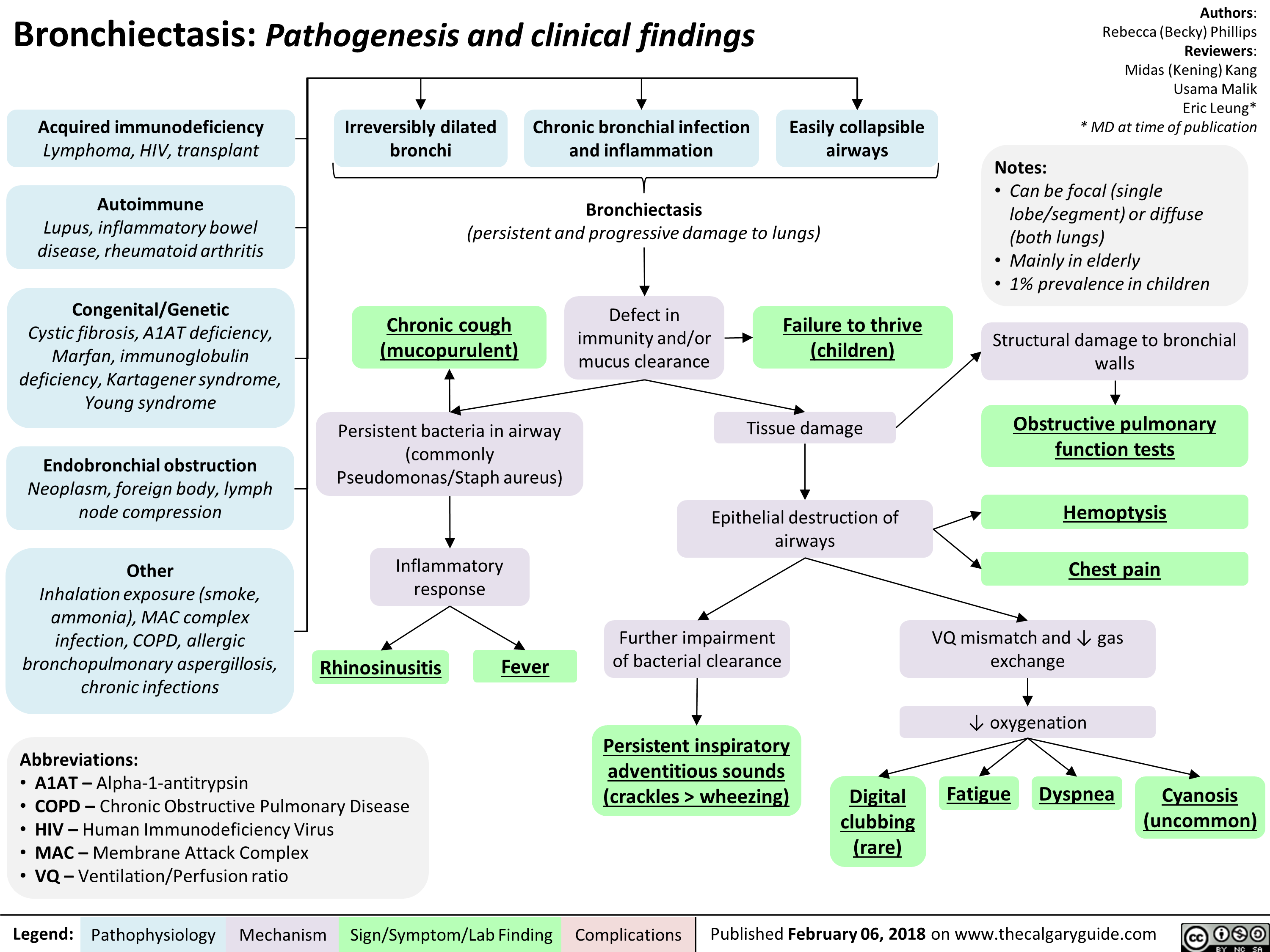
Bronchiectasis: Pathogenesis and clinical findings 
Acquired immunodeficiency Lymphoma, HIV, transplant 
Autoimmune Lupus, inflammatory bowel disease, rheumatoid arthritis 
Congenital/Genetic Cystic fibrosis, A1AT deficiency, Marfan, immunoglobulin deficiency, Kartagener syndrome, Young syndrome 
Endobronchial obstruction Neoplasm, foreign body, lymph node compression 
Other Inhalation exposure (smoke, ammonia), MAC complex infection, COPD, allergic bronchopulmonary aspergillosis, chronic infections 
Irreversibly dilated bronchi 
Chronic bronchial infection and inflammation 
1 
Easily collapsible airways 
 I Bronchiectasis (persistent and progressive damage to lungs) 
Chronic cough  (mucopurulent) 
Defect in immunity and/or mucus clearance 
Persistent bacteria in airway (commonly Pseudomonas/Staph aureus) 
Inflammatory response 
Rhinosinusitis 
Abbreviations: • A1AT — Alpha-1-antitrypsin • COPD — Chronic Obstructive Pulmonary Disease • HIV — Human Immunodeficiency Virus • MAC — Membrane Attack Complex • VQ— Ventilation/Perfusion ratio 
Legend: 
Pathophysiology Mechanism 
Fever 
Sign/Symptom/Lab Finding 
Failure to thrive (children)  

Authors: Rebecca (Becky) Phillips Reviewers: Midas (Kening) Kang Usama Malik Eric Leung* * MD at time of publication 
Notes: • Can be focal (single lobe/segment) or diffuse (both lungs) • Mainly in elderly • 1% prevalence in children 
Tissue damage 
Epithelial destruction of airways 
Further impairment of bacterial clearance 
Persistent inspiratory adventitious sounds  (crackles > wheezing)  
Complications 
Structural damage to bronchial walls 
Obstructive pulmonary function tests  
Hemoptysis 
Chest pain 
VQ mismatch and 4, gas exchange 
4, oxygenation 

Digital  clubbing (rare)  
Fatigue Dyspnea  
Cyanosis  (uncommon)

Orbital Cellulitis: Pathogenesis and clinical findings
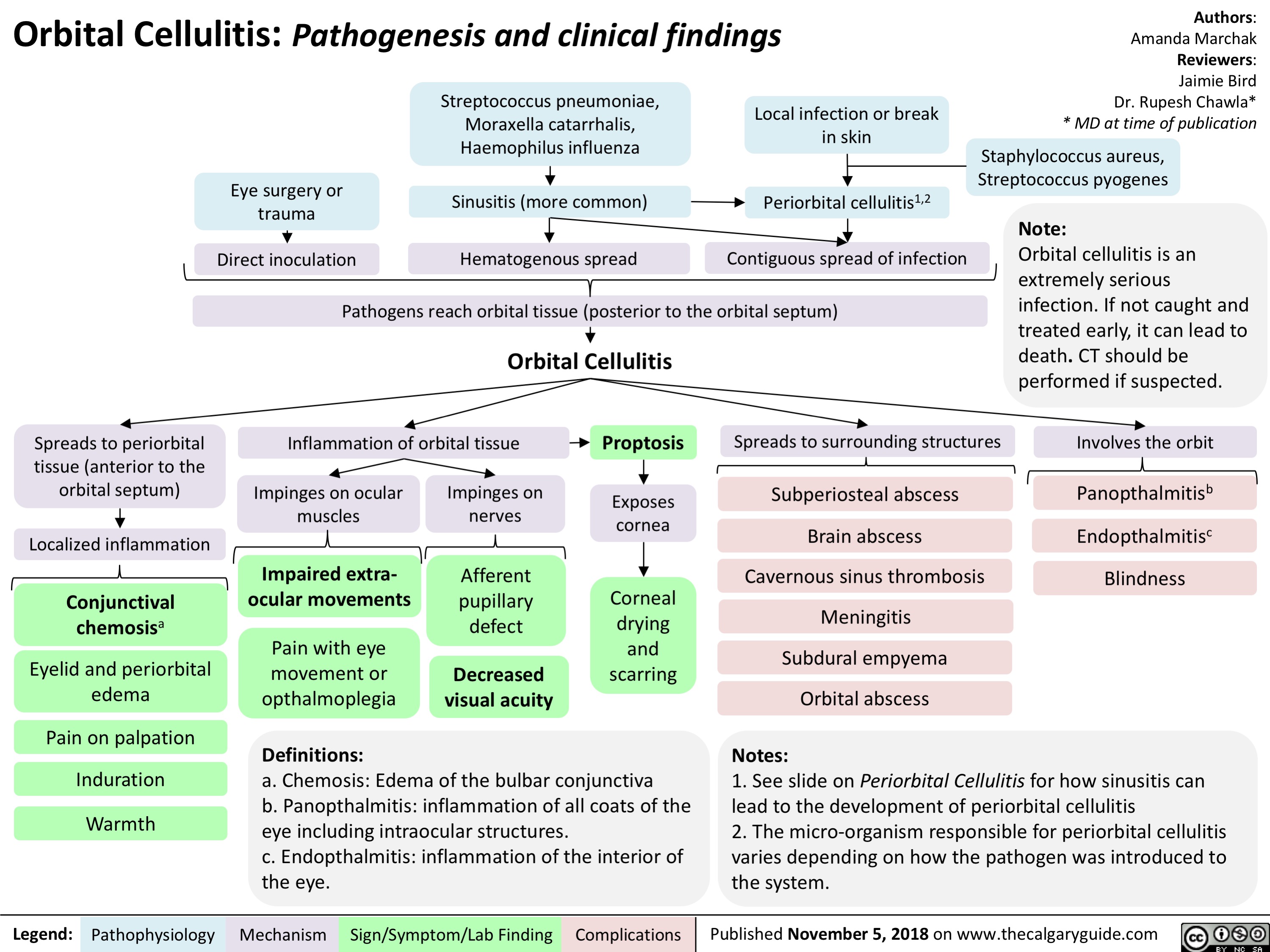
Orbital Cellulitis: Pathogenesis and clinical findings
Authors: Amanda Marchak Reviewers: Jaimie Bird Dr. Rupesh Chawla* * MD at time of publication
Staphylococcus aureus, Streptococcus pyogenes
Note:
Orbital cellulitis is an extremely serious infection. If not caught and treated early, it can lead to death. CT should be performed if suspected.
Involves the orbit
Panopthalmitisb Endopthalmitisc Blindness
 Streptococcus pneumoniae, Moraxella catarrhalis, Haemophilus influenza
Local infection or break in skin
      Eye surgery or trauma
Direct inoculation
Sinusitis (more common)       Periorbital cellulitis1,2
       Hematogenous spread
Contiguous spread of infection
  Pathogens reach orbital tissue (posterior to the orbital septum)
        Spreads to periorbital tissue (anterior to the orbital septum)
Localized inflammation
Conjunctival chemosisa
Eyelid and periorbital edema
Pain on palpation
Induration
Warmth
Orbital Cellulitis Inflammation of orbital tissue       Proptosis
Spreads to surrounding structures
Subperiosteal abscess Brain abscess Cavernous sinus thrombosis Meningitis Subdural empyema Orbital abscess
Notes:
        Impinges on ocular muscles
Impaired extra- ocular movements
Pain with eye
movement or opthalmoplegia
Definitions:
Impinges on nerves
Afferent pupillary defect
Decreased visual acuity
Exposes cornea
Corneal drying and scarring
                         a. Chemosis: Edema of the bulbar conjunctiva
b. Panopthalmitis: inflammation of all coats of the eye including intraocular structures.
c. Endopthalmitis: inflammation of the interior of the eye.
1. See slide on Periorbital Cellulitis for how sinusitis can lead to the development of periorbital cellulitis
2. The micro-organism responsible for periorbital cellulitis varies depending on how the pathogen was introduced to the system.
  Legend:
 Pathophysiology
 Mechanism
Sign/Symptom/Lab Finding
  Complications
Published November 5, 2018 on www.thecalgaryguide.com

Periorbital Cellulitis: Pathogenesis and Clinical Findings
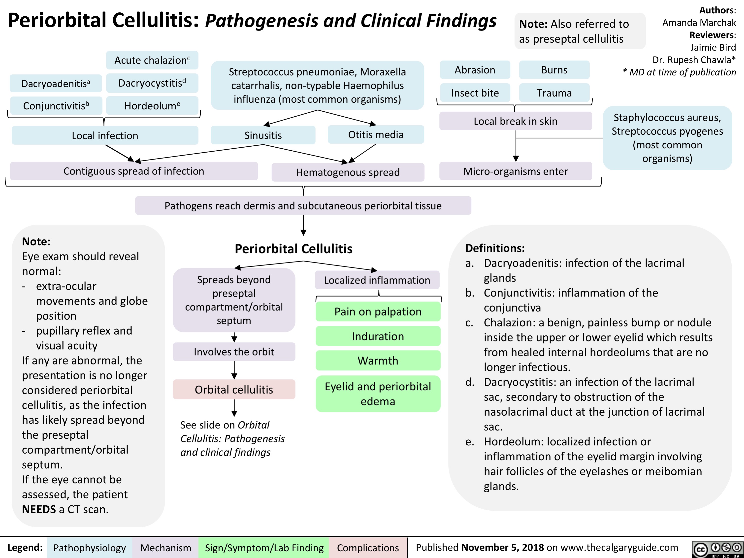
Periorbital Cellulitis: Pathogenesis and Clinical Findings
Authors: Amanda Marchak Reviewers: Jaimie Bird Dr. Rupesh Chawla* * MD at time of publication
Staphylococcus aureus, Streptococcus pyogenes (most common organisms)
 Note: Also referred to as preseptal cellulitis
      Dacryoadenitisa Conjunctivitisb
Acute chalazionc
Dacryocystitisd Hordeolume
Streptococcus pneumoniae, Moraxella catarrhalis, non-typable Haemophilus influenza (most common organisms)
Abrasion Insect bite
Burns Trauma
             Local infection
Contiguous spread of infection
Sinusitis
Otitis media Hematogenous spread
Local break in skin Micro-organisms enter
Definitions:
              Note:
Eye exam should reveal normal:
- extra-ocular
movements and globe
position
- pupillary reflex and
visual acuity
If any are abnormal, the presentation is no longer considered periorbital cellulitis, as the infection has likely spread beyond the preseptal compartment/orbital septum.
If the eye cannot be assessed, the patient NEEDS a CT scan.
Pathogens reach dermis and subcutaneous periorbital tissue
Periorbital Cellulitis
a. Dacryoadenitis: infection of the lacrimal glands
b. Conjunctivitis: inflammation of the conjunctiva
c. Chalazion: a benign, painless bump or nodule inside the upper or lower eyelid which results from healed internal hordeolums that are no longer infectious.
d. Dacryocystitis: an infection of the lacrimal sac, secondary to obstruction of the nasolacrimal duct at the junction of lacrimal sac.
e. Hordeolum: localized infection or inflammation of the eyelid margin involving hair follicles of the eyelashes or meibomian glands.
   Spreads beyond preseptal compartment/orbital septum
Involves the orbit Orbital cellulitis
See slide on Orbital Cellulitis: Pathogenesis and clinical findings
Localized inflammation
Pain on palpation
Induration
Warmth
Eyelid and periorbital edema
           Legend:
 Pathophysiology
 Mechanism
Sign/Symptom/Lab Finding
  Complications
Published November 5, 2018 on www.thecalgaryguide.com

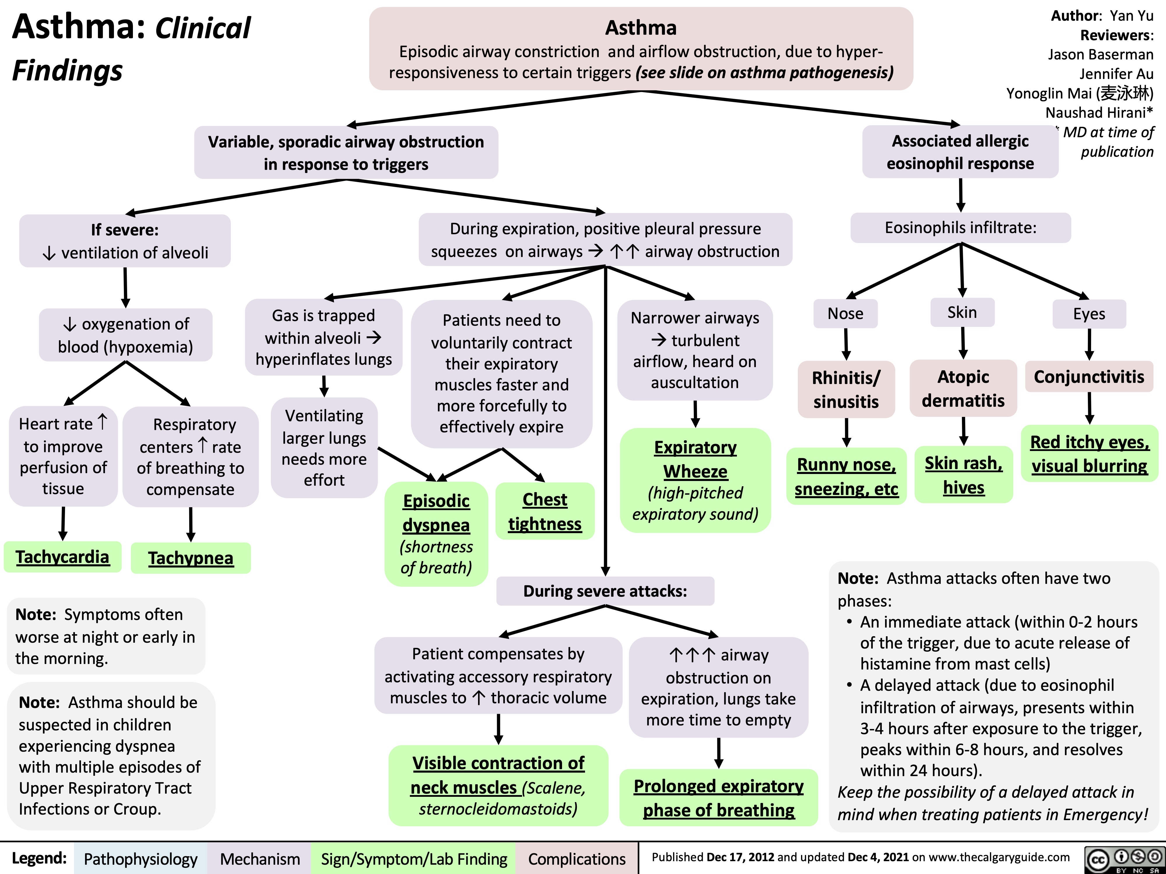
Asthma clinical findings

Asthma: Clinical Findings
Asthma
Episodic airway constriction and airflow obstruction, due to hyper- responsiveness to certain triggers (see slide on asthma pathogenesis)
Author: Yan Yu Reviewers: Jason Baserman Jennifer Au Yonoglin Mai (麦泳琳) Naushad Hirani* * MD at time of publication
     Variable, sporadic airway obstruction in response to triggers
Associated allergic eosinophil response
Eosinophils infiltrate: Skin
    If severe:
↓ ventilation of alveoli
↓ oxygenation of blood (hypoxemia)
During expiration, positive pleural pressure squeezes on airwaysà↑↑ airway obstruction
                     Heart rate ­ to improve
perfusion of tissue
Tachycardia
Respiratory centers ­ rate of breathing to
compensate
Tachypnea
Gas is trapped within alveolià hyperinflates lungs
Ventilating larger lungs needs more effort
Patients need to voluntarily contract
their expiratory muscles faster and more forcefully to effectively expire
Narrower airways àturbulent
airflow, heard on auscultation
Expiratory Wheeze (high-pitched expiratory sound)
Nose
Rhinitis/ sinusitis
Runny nose, sneezing, etc
Atopic dermatitis
Skin rash, hives
Eyes
Conjunctivitis
Red itchy eyes, visual blurring
        Episodic
dyspnea
(shortness of breath)
Chest tightness
       During severe attacks:
Note: Asthma attacks often have two phases:
• An immediate attack (within 0-2 hours of the trigger, due to acute release of histamine from mast cells)
• A delayed attack (due to eosinophil infiltration of airways, presents within 3-4 hours after exposure to the trigger, peaks within 6-8 hours, and resolves within 24 hours).
Keep the possibility of a delayed attack in mind when treating patients in Emergency!
 Note: Symptoms often worse at night or early in the morning.
Note: Asthma should be suspected in children experiencing dyspnea with multiple episodes of Upper Respiratory Tract Infections or Croup.
Patient compensates by activating accessory respiratory muscles to ↑ thoracic volume
Visible contraction of
neck muscles (Scalene, sternocleidomastoids)
↑↑↑ airway obstruction on
expiration, lungs take more time to empty
Prolonged expiratory phase of breathing
        Legend:
 Pathophysiology
Mechanism
Sign/Symptom/Lab Finding
 Complications
 Published Dec 17, 2012 and updated Dec 4, 2021 on www.thecalgaryguide.com

Granulomatosis with Polyangiitis Pathogenesis
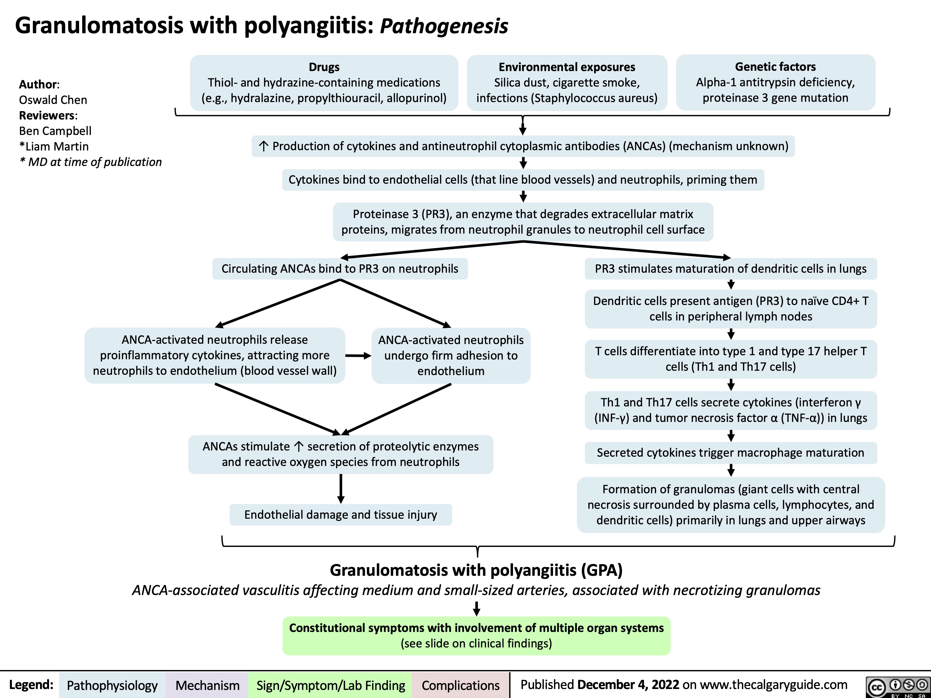
Granulomatosis with polyangiitis: Pathogenesis
   Author:
Oswald Chen
Reviewers:
Ben Campbell
*Liam Martin
* MD at time of publication
Drugs
Thiol- and hydrazine-containing medications (e.g., hydralazine, propylthiouracil, allopurinol)
Environmental exposures
Silica dust, cigarette smoke, infections (Staphylococcus aureus)
Genetic factors
Alpha-1 antitrypsin deficiency, proteinase 3 gene mutation
  ↑ Production of cytokines and antineutrophil cytoplasmic antibodies (ANCAs) (mechanism unknown) Cytokines bind to endothelial cells (that line blood vessels) and neutrophils, priming them
Proteinase 3 (PR3), an enzyme that degrades extracellular matrix proteins, migrates from neutrophil granules to neutrophil cell surface
      Circulating ANCAs bind to PR3 on neutrophils
PR3 stimulates maturation of dendritic cells in lungs
Dendritic cells present antigen (PR3) to naïve CD4+ T cells in peripheral lymph nodes
T cells differentiate into type 1 and type 17 helper T cells (Th1 and Th17 cells)
Th1 and Th17 cells secrete cytokines (interferon γ (INF-γ) and tumor necrosis factor α (TNF-α)) in lungs
Secreted cytokines trigger macrophage maturation
Formation of granulomas (giant cells with central necrosis surrounded by plasma cells, lymphocytes, and dendritic cells) primarily in lungs and upper airways
    ANCA-activated neutrophils release proinflammatory cytokines, attracting more neutrophils to endothelium (blood vessel wall)
ANCA-activated neutrophils undergo firm adhesion to endothelium
     ANCAs stimulate ↑ secretion of proteolytic enzymes and reactive oxygen species from neutrophils
Endothelial damage and tissue injury
    Granulomatosis with polyangiitis (GPA)
ANCA-associated vasculitis affecting medium and small-sized arteries, associated with necrotizing granulomas
Constitutional symptoms with involvement of multiple organ systems
 (see slide on clinical findings)
 Legend:
 Pathophysiology
Mechanism
Sign/Symptom/Lab Finding
 Complications
Published December 4, 2022 on www.thecalgaryguide.com
    
Granulomatosis with polyangiitis: Clinical findings Inflammation-mediated endothelial damage and granuloma formation
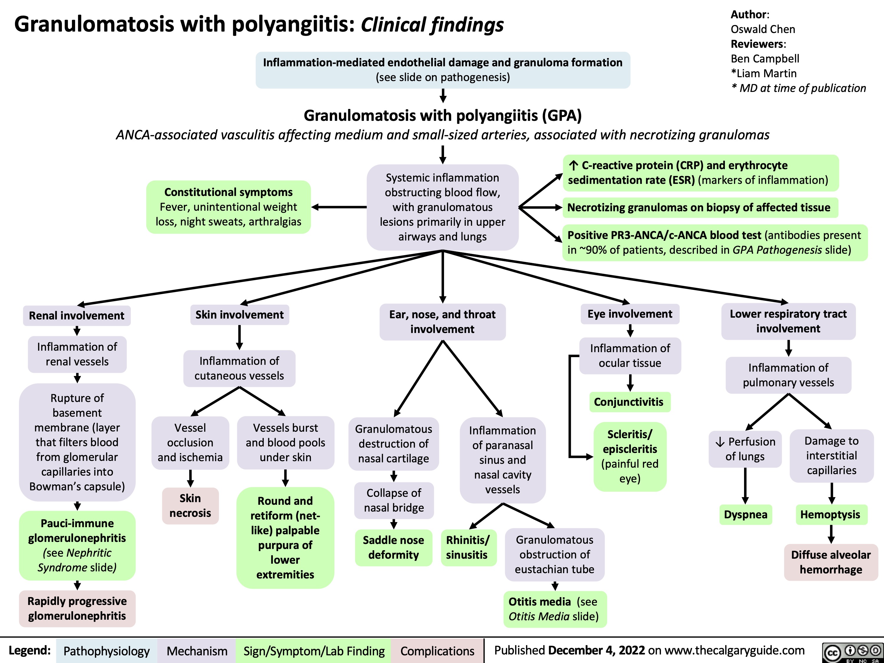
(see slide on pathogenesis)
Granulomatosis with polyangiitis (GPA)
Author:
Oswald Chen
Reviewers:
Ben Campbell
*Liam Martin
* MD at time of publication
 ANCA-associated vasculitis affecting medium and small-sized arteries, associated with necrotizing granulomas
    Constitutional symptoms
Fever, unintentional weight loss, night sweats, arthralgias
Skin involvement
Inflammation of cutaneous vessels
Systemic inflammation obstructing blood flow, with granulomatous lesions primarily in upper airways and lungs
Ear, nose, and throat involvement
↑ C-reactive protein (CRP) and erythrocyte sedimentation rate (ESR) (markers of inflammation)
Necrotizing granulomas on biopsy of affected tissue
Positive PR3-ANCA/c-ANCA blood test (antibodies present in ~90% of patients, described in GPA Pathogenesis slide)
             Renal involvement
Inflammation of renal vessels
Rupture of basement membrane (layer that filters blood from glomerular capillaries into Bowman’s capsule)
Pauci-immune glomerulonephritis (see Nephritic Syndrome slide)
Rapidly progressive glomerulonephritis
Eye involvement
Inflammation of ocular tissue
Conjunctivitis
Scleritis/ episcleritis (painful red eye)
Lower respiratory tract involvement
Inflammation of pulmonary vessels
                 Vessel occlusion and ischemia
Skin necrosis
Vessels burst and blood pools under skin
Round and retiform (net- like) palpable purpura of lower extremities
Granulomatous destruction of nasal cartilage
Collapse of nasal bridge
Saddle nose deformity
Inflammation of paranasal sinus and nasal cavity vessels
↓ Perfusion of lungs
Dyspnea
Damage to interstitial capillaries
Hemoptysis
Diffuse alveolar hemorrhage
              Rhinitis/ sinusitis
Granulomatous obstruction of eustachian tube
Otitis media (see Otitis Media slide)
    Legend:
 Pathophysiology
Mechanism
Sign/Symptom/Lab Finding
 Complications
Published December 4, 2022 on www.thecalgaryguide.com

Granulomatosis with polyangiitis: Clinical findings

Granulomatosis with polyangiitis: Clinical findings Inflammation-mediated endothelial damage and granuloma formation
(see slide on pathogenesis)
Granulomatosis with polyangiitis (GPA)
Author:
Oswald Chen
Reviewers:
Ben Campbell
*Liam Martin
* MD at time of publication
 ANCA-associated vasculitis affecting medium and small-sized arteries, associated with necrotizing granulomas
    Constitutional symptoms
Fever, unintentional weight loss, night sweats, arthralgias
Skin involvement
Inflammation of cutaneous vessels
Systemic inflammation obstructing blood flow, with granulomatous lesions primarily in upper airways and lungs
Ear, nose, and throat involvement
↑ C-reactive protein (CRP) and erythrocyte sedimentation rate (ESR) (markers of inflammation)
Necrotizing granulomas on biopsy of affected tissue
Positive PR3-ANCA/c-ANCA blood test (antibodies present in ~90% of patients, described in GPA Pathogenesis slide)
             Renal involvement
Inflammation of renal vessels
Rupture of basement membrane (layer that filters blood from glomerular capillaries into Bowman’s capsule)
Pauci-immune glomerulonephritis (see Nephritic Syndrome slide)
Rapidly progressive glomerulonephritis
Eye involvement
Inflammation of ocular tissue
Conjunctivitis
Scleritis/ episcleritis (painful red eye)
Lower respiratory tract involvement
Inflammation of pulmonary vessels
                 Vessel occlusion and ischemia
Skin necrosis
Vessels burst and blood pools under skin
Round and retiform (net- like) palpable purpura of lower extremities
Granulomatous destruction of nasal cartilage
Collapse of nasal bridge
Saddle nose deformity
Inflammation of paranasal sinus and nasal cavity vessels
↓ Perfusion of lungs
Dyspnea
Damage to interstitial capillaries
Hemoptysis
Diffuse alveolar hemorrhage
              Rhinitis/ sinusitis
Granulomatous obstruction of eustachian tube
Otitis media (see Otitis Media slide)
   
Legend:
Pathophysiology
Mechanism
Sign/Symptom/Lab Finding
Complications
Published December 4, 2022 on www.thecalgaryguide.com

Tonsillitis Pathogenesis and clinical findings
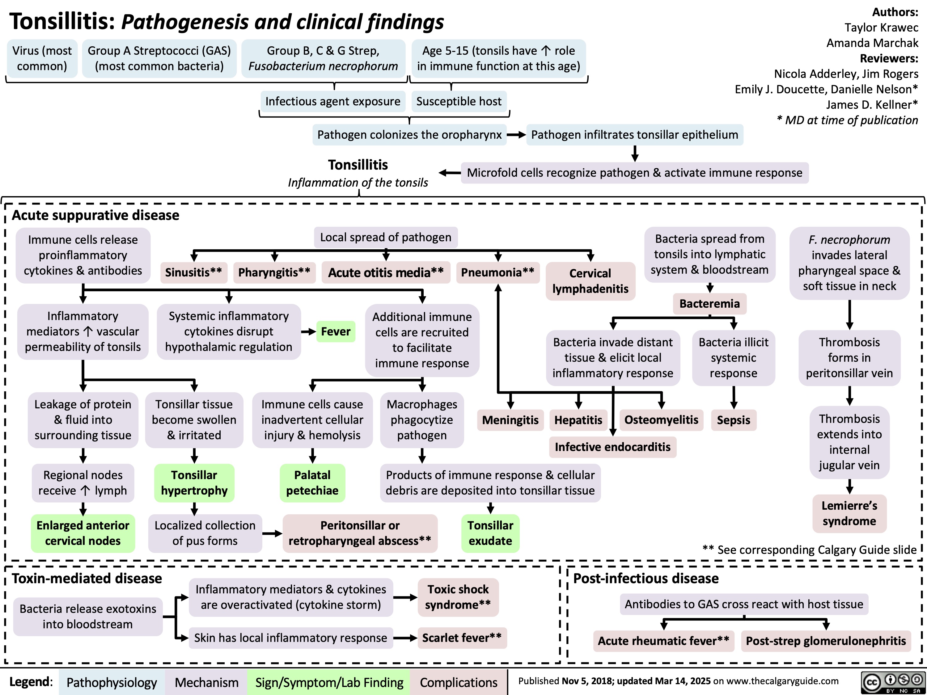
Tonsillitis: Pathogenesis and clinical findings
Authors:
Taylor Krawec Amanda Marchak Reviewers: Nicola Adderley, Jim Rogers Emily J. Doucette, Danielle Nelson* James D. Kellner* * MD at time of publication
Pathogen infiltrates tonsillar epithelium
Microfold cells recognize pathogen & activate immune response
    Virus (most common)
Group A Streptococci (GAS) (most common bacteria)
Group B, C & G Strep,
Fusobacterium necrophorum
Age 5-15 (tonsils have ↑ role in immune function at this age)
Tonsillitis
Inflammation of the tonsils
    Infectious agent exposure
Susceptible host Pathogen colonizes the oropharynx
     Acute suppurative disease
    Immune cells release proinflammatory cytokines & antibodies
Inflammatory mediators ↑ vascular permeability of tonsils
Leakage of protein & fluid into surrounding tissue
Regional nodes receive ↑ lymph
Enlarged anterior cervical nodes
Sinusitis**
Pharyngitis**
Local spread of pathogen
Acute otitis media**
Pneumonia**
Cervical lymphadenitis
Bacteria spread from
tonsils into lymphatic system & bloodstream
Bacteremia
F. necrophorum
invades lateral pharyngeal space & soft tissue in neck
Thrombosis forms in peritonsillar vein
Thrombosis extends into internal jugular vein
Lemierre’s syndrome
               Systemic inflammatory cytokines disrupt hypothalamic regulation
Fever
Additional immune cells are recruited to facilitate immune response
Macrophages phagocytize pathogen
Bacteria invade distant tissue & elicit local inflammatory response
Hepatitis Osteomyelitis
Infective endocarditis
Bacteria illicit systemic response
Sepsis
           Tonsillar tissue become swollen & irritated
Tonsillar hypertrophy
Localized collection of pus forms
Immune cells cause inadvertent cellular injury & hemolysis
Palatal petechiae
Meningitis
        Products of immune response & cellular debris are deposited into tonsillar tissue
     Peritonsillar or Tonsillar retropharyngeal abscess** exudate
** See corresponding Calgary Guide slide
 Toxin-mediated disease
Bacteria release exotoxins into bloodstream
Inflammatory mediators & cytokines are overactivated (cytokine storm)
Skin has local inflammatory response
Toxic shock syndrome**
     Scarlet fever**
 Post-infectious disease
Antibodies to GAS cross react with host tissue
     Acute rheumatic fever** Post-strep glomerulonephritis
Legend:
 Pathophysiology
 Mechanism
Sign/Symptom/Lab Finding
 Complications
 Published Nov 5, 2018; updated Mar 14, 2025 on www.thecalgaryguide.com

Cystic Fibrosis

Authors:
Navdeep Goraya, Spencer Montgomery
Reviewers:
Yan Yu, Kayla Nelson, Emily J. Doucette,
Mark Montgomery*, Danielle Nelson*
*MD at time of publication
Reproductive Manifestations
Incomplete development of Wolffian
duct derivatives (vas deferens,
epididymis, & seminal vesicles)
Cystic Fibrosis (CF): Pathogenesis, clinical findings, and complications
Cystic Fibrosis Transmembrane Regulator (CFTR) autosomal recessive gene mutation on chromosome 7
CFTR protein (transmembrane chloride ion
channel found in exocrine tissue) dysfunction
Mutated CFTR
proteins prevent
Cl- reabsorption
in sweat glands
↑ Secretion of
Cl- into sweat
↑ Sweat Cl-
concentration
Mutated CFTR proteins in duct epithelial
tissue of other parts of the body prevent
diffusion of Cl- into secretions
↓ Cl- diffusion into peri-ciliary fluid
↓ Water composition of peri-ciliary fluid
↓ Clearance of mucociliary secretions
Secretions accumulate in secretory
passages throughout the body
Inhibition of sperm transport
(obstructive azoospermia)
Male
infertility
Upper Respiratory Tract Manifestations
Retained secretions
in sinuses
Failure to clear
bacteria in sinuses
Persistent neutrophilic inflammation triggers
tissue remodeling & mucosal overgrowth
Bacterial
proliferation
Nasal
polyps
Chronic
sinusitis
Pancreatic Manifestations
Trapped digestive
enzymes degrade
pancreatic tissue
Pancreatic tissue
damage triggers
inflammation,
scarring & fatty
tissue replacement
Islet cell damage
& destruction
Cystic-fibrosis related
diabetes (CFRD)
Lower Respiratory Tract Manifestations
Retained secretions
in airways
Bacterial proliferation
in lower airway
Airway infection
& inflammation
Chronic
productive cough
Signs of obstructive lung disease (lung hyperinflation
on x-ray & abnormal pulmonary function tests)
Bronchitis ±
bronchiectasis**
↓ Production & secretion of
pancreatic enzymes into GI
tract (pancreatic insufficiency)
Fat & protein malabsorption
Failure to
thrive
↓ Absorption of
fat-soluble vitamins
Steatorrhea
(↑ fat in stool)
Vitamin D
deficiency
Vitamin K
deficiency**
Rickets**
Osteoporosis**
Coagulopathies
Hepatic Manifestations
Delayed passage of bile
through biliary tree
↑ Loss of bile acids in stool
Inflammatory hepatic
response
↑ Production of lithogenic bile (bile
supersaturated with cholesterol)
Biliary cirrhosis with
portal hypertension
Cholelithiasis**
Gastrointestinal (GI) Manifestations
↓ Movement of
intestinal contents
In newborns:
Meconium ileus
In children/adults: Distal ileal
obstruction syndrome (DIOS)
↑ Retention
of meconium
↑ Reabsorption
of bilirubin
Prolonged jaundice
in neonates
**See corresponding Calgary Guide slide
Legend: Sign/Symptom/Lab Finding Complications
Pathophysiology Mechanism
Published Jan 21, 2013; updated Aug 20, 2025 on www.thecalgaryguide.com
Reproductive Manifestations
Degeneration of Wolffian duct derivatives
(vas deferens, epididymis, & seminal vesicles)
Inhibition of sperm transport
(obstructive azoospermia)
Male
infertility
Cystic Fibrosis (CF): Pathogenesis, clinical findings, and complications
Cystic Fibrosis Transmembrane Regulator (CFTR) autosomal recessive gene mutation on chromosome 7
CFTR protein (a transmembrane chloride ion
channel that is found in exocrine tissue) dysfunction
Authors:
Navdeep Goraya, Spencer Montgomery
Reviewers:
Yan Yu, Kayla Nelson, Emily J. Doucette,
Mark Montgomery*, Danielle Nelson*
*MD at time of publication
Mutated CFTR
proteins prevent
Cl- reabsorption
in sweat glands
↑ Secretion of
Cl- into sweat
↑ Sweat Cl-
concentration
Mutated CFTR proteins in duct epithelial
tissue of other parts of the body prevent
diffusion of Cl- into secretions
↓ Cl- diffusion into peri-ciliary fluid
↓ Water composition of peri-ciliary fluid
↓ Clearance of
mucociliary secretions
Secretions accumulate in secretory
passages throughout the body
Upper Respiratory Tract Manifestations
Retained secretions
in sinuses
Failure to clear
bacteria in sinuses
Persistent neutrophilic inflammation triggers
tissue remodeling & mucosal overgrowth
Bacterial
proliferation
Nasal
polyps
Chronic
sinusitis
Lower Respiratory Tract Manifestations
Retained secretions
in airways
Bacterial proliferation
in lower airway
Airway infection
& inflammation
Chronic
productive cough
Signs of obstructive lung disease (lung hyperinflation
on x-ray & abnormal pulmonary function tests)
Bronchitis ±
bronchiectasis**
Pancreatic Manifestations
Pancreas unable to
secrete digestive enzymes
into GI tract (pancreatic
insufficiency)
Fat & protein
malabsorption
↓ Absorption of
fat-soluble vitamins
Failure to
thrive
↓ Serum Vitamin D
Osteoporosis**
Trapped digestive
enzymes degrade
pancreatic tissue
Tissue damage
triggers inflammation,
scarring & fatty tissue
replacement
Islet cell
destruction
Cystic-fibrosis related
diabetes (CFRD)
Hepatic Manifestations
Delayed passage of bile
through biliary tree
Inflammatory hepatic
response
Cirrhosis** & portal
hypertension
Gastrointestinal Manifestations
↓ Movement of
intestinal contents
In newborns:
Meconium ileus
In children/adults: Distal ileal
obstruction syndrome (DIOS)
↑ Retention
of meconium
↑ Reabsorption
of bilirubin
Prolonged jaundice
in neonates
Legend: Pathophysiology Mechanism
Sign/Symptom/Lab Finding Complications
**See corresponding Calgary Guide slide
Published January 21, 2013 on www.thecalgaryguide.com
Please only review slide 1 – slides 3-7 are previous draft
versions.
Thank you!
Authors:
Spencer Montgomery, Navdeep Goraya
Reviewers:
Yan Yu, Kayla Nelson, Emily J. Doucette,
Mark Montgomery*, Name Name*
*MD at time of publication
In the vas deferens
in utero
Cystic Fibrosis: Pathogenesis, clinical findings, and complications
Cystic Fibrosis Transmembrane Regulator (CFTR) autosomal recessive gene mutation on chromosome 7
CFTR protein (a transmembrane chloride ion
channel that is found in exocrine tissue) dysfunction
Mutated CFTR
proteins prevent
Cl- reabsorption
in sweat glands
↑ Secretion of
Cl- into sweat
↑ Sweat Cl-
concentration
Mutated CFTR proteins in duct epithelial
tissue of other parts of the body prevent
diffusion of Cl- into secretions
↓ Cl- diffusion into peri-ciliary fluid
↓ Water composition of peri-ciliary fluid
↓ Clearance of
mucociliary secretions
Secretions accumulate in secretory
passages throughout the body
Degeneration of vas deferens, Wolffian
ducts & associated structures
Infertility in
affected males
In upper
respiratory
tract
Retained
secretions
in sinuses
Failure to clear
bacteria in
airways
Persistent neutrophilic inflammation triggers
tissue remodeling & mucosal overgrowth
Bacterial
proliferation
Chronic
sinusitis
Nasal polyps
In lower
respiratory
tract
Chronic
productive cough
Retained
secretions in
airways
Bacterial
proliferation
Airway
infection &
inflammation
Signs of obstructive lung disease (lung hyperinflation
on x-ray & abnormal pulmonary function tests)
Bronchitis ±
bronchiectasis**
In pancreas
Pancreas unable to secrete
digestive enzymes into GI tract
(pancreatic insufficiency)
Fat & protein
malabsorption
↓ Absorption of fat-
soluble vitamins
Failure to
thrive
↓ Serum Vitamin D
Osteoporosis**
Trapped digestive
enzymes degrade
pancreatic tissue
Tissue damage triggers
inflammation, scarring
& fatty tissue
replacement
Islet cell
destruction
Cystic-fibrosis related
diabetes (CFRD)
In biliary tree
Delayed
passage of bile
Inflammatory hepatic
response
Cirrhosis** &
portal
hypertension
In GI tract
↓ Movement
of intestinal
contents
In children/adults: Distal ileal
obstruction syndrome (DIOS)
In newborns:
Meconium
ileus
↑ Retention
of meconium
↑ Reabsorption
of bilirubin
Prolonged
jaundice in
neonates
Legend: Pathophysiology Mechanism
Sign/Symptom/Lab Finding Complications
Published January 21, 2013 on www.thecalgaryguide.com
Authors:
Spencer Montgomery, Navdeep Goraya
Reviewers:
Yan Yu, Kayla Nelson, Emily J. Doucette,
Mark Montgomery*, Name Name*
*MD at time of publication
In the vas deferens
in utero
Cystic Fibrosis: Pathogenesis, clinical findings, and complications
Cystic Fibrosis Transmembrane Regulator (CFTR) autosomal recessive gene mutation on chromosome 7
CFTR protein (a transmembrane chloride ion
channel that is found in exocrine tissue) dysfunction
Mutated CFTR
proteins prevent
Cl- reabsorption
in sweat glands
↑ Secretion of
Cl- into sweat
↑ Sweat Cl-
concentration
Mutated CFTR proteins in duct epithelial
tissue of other parts of the body prevent
diffusion of Cl- into secretions
↓ Cl- diffusion into peri-ciliary fluid
↓ Water composition of peri-ciliary fluid
↓ Clearance of
mucociliary secretions
Secretions accumulate in secretory
passages throughout the body
Degeneration of vas deferens, Wolffian
ducts & associated structures
Infertility in
affected males
In upper
respiratory
tract
Retained
secretions
in sinuses
Failure to clear
bacteria in
airways
Persistent neutrophilic inflammation triggers
tissue remodeling & mucosal overgrowth
Bacterial
proliferation
Chronic
sinusitis
Nasal polyps
In lower
respiratory
tract
Chronic
productive cough
Retained
secretions in
airways
Bacterial
proliferation
Airway
infection &
inflammation
Signs of obstructive lung disease (lung hyperinflation
on x-ray & abnormal pulmonary function tests)
Bronchitis ±
bronchiectasis**
Trapped digestive
enzymes degrade
pancreatic tissue
Inflammation
Scarring & fatty
tissue infiltration
Islet cell
destruction
Type II Diabetes
Mellitus**
In pancreas
Pancreas unable to secrete
digestive enzymes into GI tract
(pancreatic insufficiency)
Fat & protein
malabsorption
↓ Absorption of fat-
soluble vitamins
Failure to
thrive
↓ Serum Vitamin D
Osteoporosis**
In biliary tree
Delayed
passage of bile
Inflammatory hepatic
response
Cirrhosis** &
portal
hypertension
In GI tract
↓ Movement
of intestinal
contents
In children/adults: Distal ileal
obstruction syndrome (DIOS)
In newborns:
Meconium
ileus
↑ Retention
of meconium
↑ Reabsorption
of bilirubin
Prolonged
jaundice in
neonates
Legend: Pathophysiology Mechanism
Sign/Symptom/Lab Finding Complications
Published January 21, 2013 on www.thecalgaryguide.com
In the vas deferens
in utero
Retained secretions
in sinuses
Cystic Fibrosis: Pathogenesis, clinical findings, and complications
Cystic Fibrosis Transmembrane Regulator (CFTR) autosomal recessive gene mutation on chromosome 7
CFTR protein (a transmembrane chloride ion
channel that is found in exocrine tissue) dysfunction
Mutated CFTR
proteins prevent
Cl- reabsorption
in sweat glands
↑ Secretion of
Cl- into sweat
↑ Sweat Cl-
concentration
Mutated CFTR proteins in duct epithelial
tissue of other parts of the body prevent
diffusion of Cl- into secretions
↓ Cl- diffusion into peri-ciliary fluid
↓ Water composition of peri-ciliary fluid
↓ Clearance of
mucociliary secretions
Accumulation of secretions in
secretory passages throughout the
body obstructing these passages
Authors:
Spencer Montgomery, Navdeep Goraya
Reviewers:
Yan Yu, Kayla Nelson,
Emily J. Doucette, Mark Montgomery*
*MD at time of publication
Degeneration of vas deferens, Wolffian
ducts & associated structures
Infertility in
affected males
In upper
respiratory
tract
Failure to clear
bacteria in
airways
Bacterial
proliferation
Chronic
sinusitis
Nasal polyps
In lower
respiratory
tract
Chronic
productive
cough
Retained
secretions in
airways
Bacterial
proliferation
Airway
infection &
inflammation
Signs of obstructive lung disease i.e. lung
hyperinflation on x-ray & abnormal pulmonary
function tests
Bronchitis ±
bronchiectasis
Trapped digestive
enzymes degrade
pancreatic tissue
Inflammation
Scarring & fatty
tissue infiltration
Islet cell
destruction
Type II Diabetes
Mellitus
In pancreas
Pancreas unable to secrete
digestive enzymes into GI tract
(pancreatic insufficiency)
Fat and protein
malabsorption
↓ Absorption of fat-
soluble vitamins
Failure to
thrive
↓Serum Vitamin D
Osteoporosis
In biliary tree
Delayed
passage
of bile
Inflammatory hepatic
response
Cirrhosis & portal
hypertension
In GI tract
↓ Movement
of intestinal
contents
In children/adults: Distal ileal
obstruction syndrome (DIOS)
In newborns:
Meconium
ileus
↑ Retention
of meconium
↑ Reabsorption
of bilirubin
Prolonged
jaundice in
neonates
Legend: Pathophysiology Mechanism
Sign/Symptom/Lab Finding Complications
Published January 21, 2013 on www.thecalgaryguide.com
In the vas
deferens
in utero
Cystic Fibrosis: Pathogenesis, clinical findings, and complications
Cystic Fibrosis Transmembrane Regulator (CFTR) autosomal recessive gene mutation on chromosome 7
CFTR protein (a transmembrane chloride ion channel that is found in
exocrine tissue) dysfunction
Chloride channel no longer allows Cl- transport
CFTR proteins in
sweat glands reabsorb
Cl-
CFTR proteins in duct epithelial tissue of
other parts of the body facilitate diffusion
of Cl- into secretions
↓Reabsorption
↓Cl- diffusion into peri-ciliary fluid
↓Water composition of peri-ciliary fluid
↑Secretion of Cl-
into sweat
↓Clearance of mucociliary secretions
↑Sweat Cl-
concentration
Accumulation of secretions in secretory
passages throughout the body obstructing
these passages
Degeneration of vas deferens, Wolffian
ducts & associated structures
Authors:
Spencer Montgomery, Navdeep Goraya
Reviewers:
Yan Yu, Kayla Nelson,
Emily J. Doucette, Mark Montgomery*
*MD at time of publication
Infertility in
affected males
In upper
respiratory
tract
Retained secretions
in sinuses
Nasal polyps
Bacterial
proliferation
Chronic
sinusitis
In lower
respiratory
tract
Chronic
productive
cough
Retained
secretions in
airways
Bacterial
proliferation
Signs of obstructive lung disease
i.e. lung hyperinflation on x-ray &
abnormal pulmonary function
tests
Airway
infection &
inflammation
Bronchitis ±
bronchiectasis
Trapped digestive
enzymes degrade
pancreatic tissue
Inflammation
Scarring & fatty
tissue infiltration
Islet cell
destruction
Type II Diabetes
Mellitus
In pancreas
Pancreas unable to secrete
digestive enzymes into GI tract
(pancreatic insufficiency)
Fat and protein
malabsorption
↓Absorption of fat-
soluble vitamins
Failure to
thrive
↓Serum Vitamin D
Osteoporosis
In biliary tree
Delayed
passage
of bile
Inflammatory hepatic
response
Cirrhosis & portal
hypertension
In GI tract
↓Movement
of intestinal
contents
In children/adults: Distal ileal obstruction
syndrome (DIOS)
In
newborns:
Meconium
ileus
↑Retention
of meconium
↑Reabsorption
of bilirubin
Prolonged
jaundice
in
neonates
Legend: Pathophysiology Mechanism
Sign/Symptom/Lab Finding Complications
Published January 21, 2013 on www.thecalgaryguide.com
In the vas
deferens
in utero
Degeneration of
vas deferens,
Wolffian ducts
and associated
structures
Infertility in
affected males
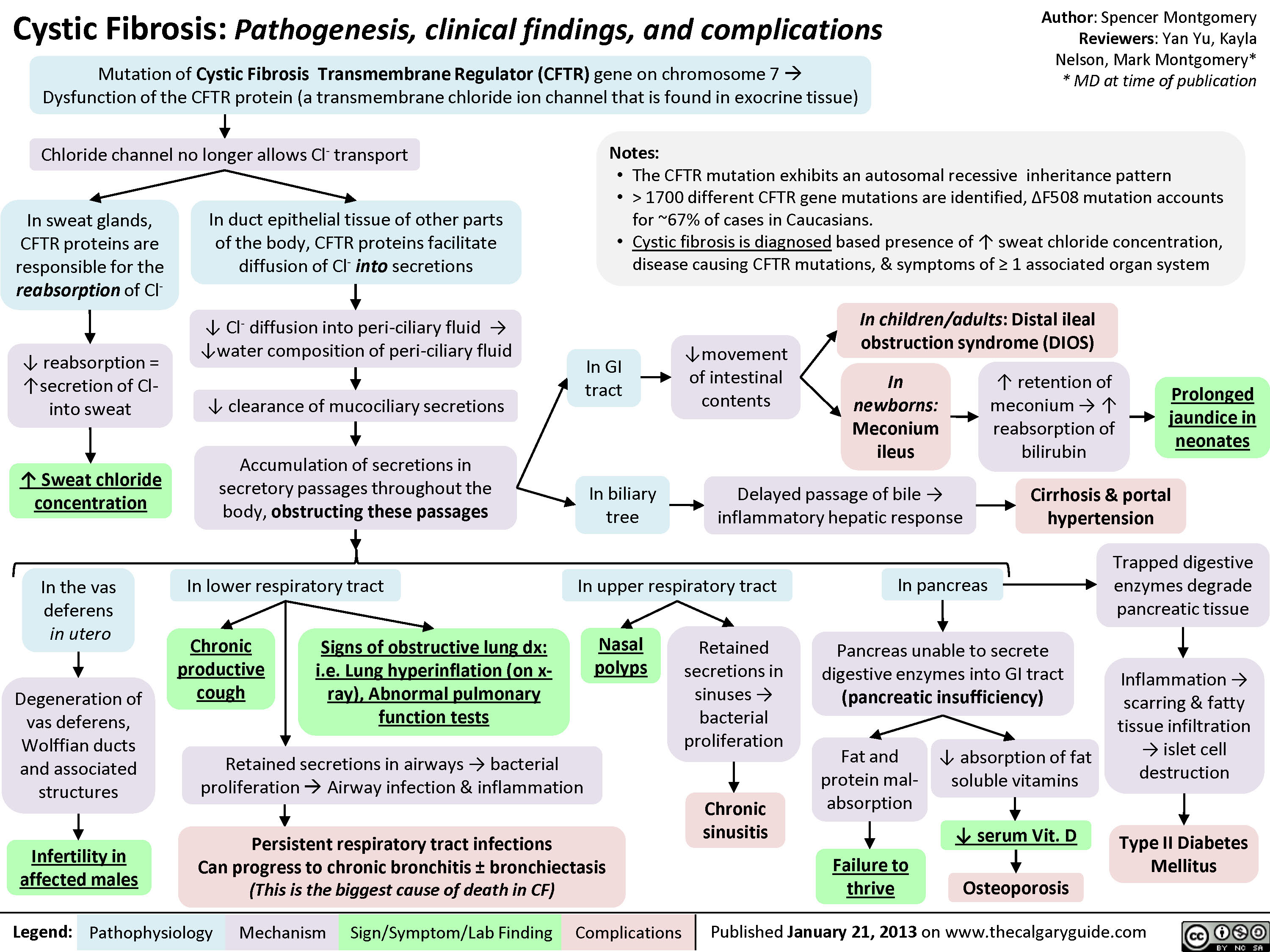
Legend: Cystic Fibrosis: Pathogenesis, clinical findings, and complications
Mutation of Cystic Fibrosis Transmembrane Regulator (CFTR) gene on chromosome 7 à
Dysfunction of the CFTR protein (a transmembrane chloride ion channel that is found in exocrine tissue)
Author: Spencer Montgomery
Reviewers: Yan Yu, Kayla
Nelson, Mark Montgomery*
* MD at time of publication
Chloride channel no longer allows Cl- transport
In sweat glands, CFTR
proteins are
responsible for the
reabsorption of Cl-
In duct epithelial tissue of other parts of
the body, CFTR proteins facilitate diffusion
of Cl- into secretions
Notes:
• The CFTR mutation exhibits an autosomal recessive inheritance pattern
• > 1700 different CFTR gene mutations are identified, ∆F508 mutation accounts for
~67% of cases in Caucasians.
• Cystic fibrosis is diagnosed based presence of ↑ sweat chloride concentration,
disease causing CFTR mutations, & symptoms of ≥ 1 associated organ system
↓ Cl- diffusion into peri-ciliary fluid →
↓water composition of peri-ciliary fluid
In children/adults: Distal ileal
obstruction syndrome (DIOS)
↓ reabsorption =
↑secretion of Cl-
into sweat
In GI
tract
↓movement
of intestinal
contents
↓ clearance of mucociliary secretions
In
newborns:
Meconium
ileus
↑ retention of
meconium → ↑
reabsorption of
bilirubin
Prolonged
jaundice in
neonates
↑ Sweat chloride
concentration
Accumulation of secretions in secretory
passages throughout the body,
obstructing these passages
In biliary
tree
Delayed passage of bile →
inflammatory hepatic response
Cirrhosis & portal
hypertension
In upper respiratory tract
In pancreas
Trapped digestive
enzymes degrade
pancreatic tissue
Nasal
polyps
Retained
secretions in
sinuses →
bacterial
proliferation
Pancreas unable to secrete
digestive enzymes into GI tract
(pancreatic insufficiency)
Fat and
protein mal-
absorption
↓ absorption of fat
soluble vitamins
Inflammation →
scarring & fatty
tissue infiltration
→ islet cell
destruction
Chronic
sinusitis
↓ serum Vit. D
Type II Diabetes
Mellitus
Failure to
thrive
Osteoporosis
Published January 21, 2013 on www.thecalgaryguide.com
In lower respiratory tract
Chronic
productive
cough
Retained secretions in airways → bacterial proliferation
à Airway infection & inflammation
Persistent respiratory tract infections
Can progress to chronic bronchitis ± bronchiectasis
(This is the biggest cause of death in CF)
Pathophysiology Signs of obstructive lung dx: i.e.
Lung hyperinflation (on x-ray),
Abnormal pulmonary function
tests
Mechanism
Sign/Symptom/Lab Finding Complications

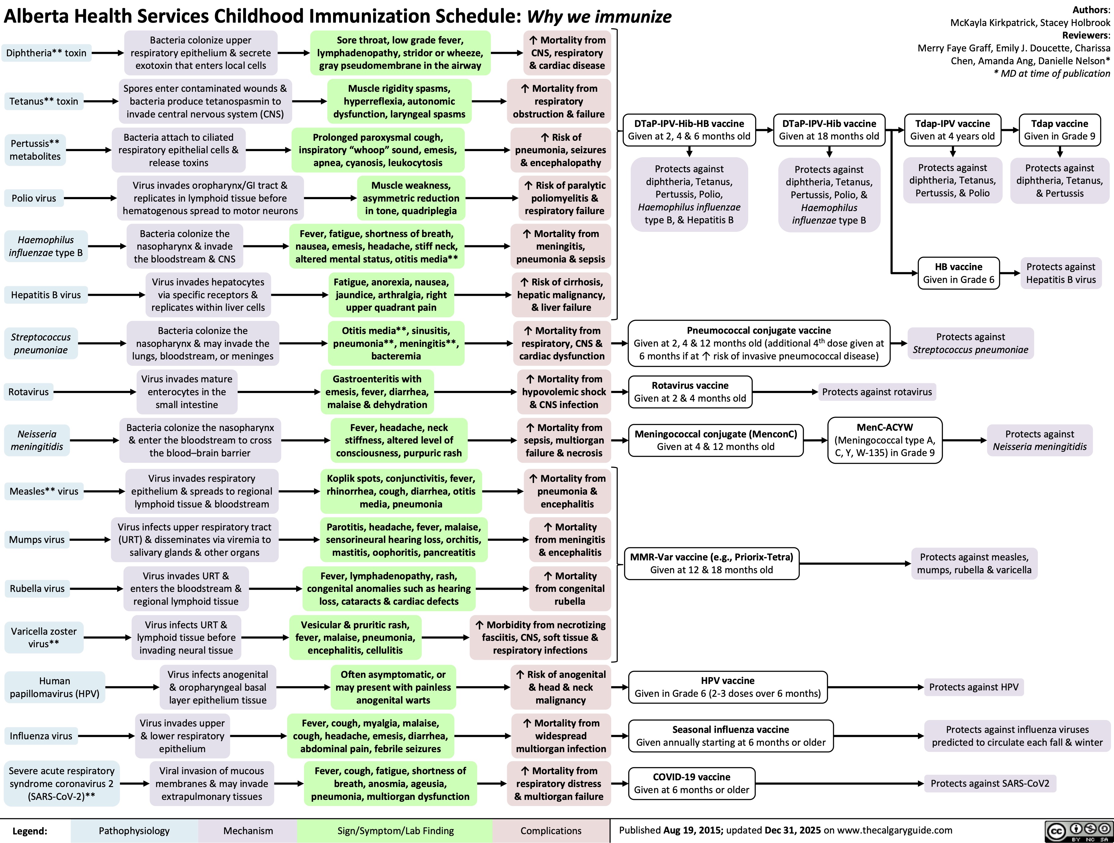
Childhood Immunization Schedule

Alberta Health Services Childhood Immunization Schedule: Why we immunize
Diphtheria** toxin
Bacteria colonize upper
respiratory epithelium & secrete
exotoxin that enters local cells
Sore throat, low grade fever,
lymphadenopathy, stridor or wheeze,
gray pseudomembrane in the airway
↑ Mortality from
CNS, respiratory
& cardiac disease
Tetanus** toxin
Pertussis**
metabolites
Polio virus
Haemophilus
influenzae type B
Hepatitis B virus
Streptococcus
pneumoniae
Rotavirus
Neisseria
meningitidis
Measles** virus
Mumps virus
Rubella virus
Varicella zoster
virus**
Human
papillomavirus (HPV)
Spores enter contaminated wounds &
bacteria produce tetanospasmin to
invade central nervous system (CNS)
Muscle rigidity spasms,
hyperreflexia, autonomic
dysfunction, laryngeal spasms
↑ Mortality from
respiratory
obstruction & failure
Bacteria attach to ciliated
respiratory epithelial cells &
release toxins
Prolonged paroxysmal cough,
inspiratory “whoop” sound, emesis,
apnea, cyanosis, leukocytosis
↑ Risk of
pneumonia, seizures
& encephalopathy
Virus invades oropharynx/GI tract &
replicates in lymphoid tissue before
hematogenous spread to motor neurons
↑ Risk of paralytic
poliomyelitis &
respiratory failure
Bacteria colonize the
nasopharynx & invade
the bloodstream & CNS
Virus invades hepatocytes
via specific receptors &
replicates within liver cells
Bacteria colonize the
nasopharynx & may invade the
lungs, bloodstream, or meninges
Virus invades mature
enterocytes in the
small intestine
Bacteria colonize the nasopharynx
& enter the bloodstream to cross
the blood–brain barrier
Virus invades respiratory
epithelium & spreads to regional
lymphoid tissue & bloodstream
Virus infects upper respiratory tract
(URT) & disseminates via viremia to
salivary glands & other organs
Virus invades URT &
enters the bloodstream &
regional lymphoid tissue
Virus infects URT &
lymphoid tissue before
invading neural tissue
Virus infects anogenital
& oropharyngeal basal
layer epithelium tissue
Influenza virus
Virus invades upper
& lower respiratory
epithelium
Severe acute respiratory
syndrome coronavirus 2
(SARS-CoV-2)**
Viral invasion of mucous
membranes & may invade
extrapulmonary tissues
Legend: Muscle weakness,
asymmetric reduction
in tone, quadriplegia
Fever, fatigue, shortness of breath,
nausea, emesis, headache, stiff neck,
altered mental status, otitis media**
Fatigue, anorexia, nausea,
jaundice, arthralgia, right
upper quadrant pain
Otitis media**, sinusitis,
pneumonia**, meningitis**,
bacteremia
Gastroenteritis with
emesis, fever, diarrhea,
malaise & dehydration
Fever, headache, neck
stiffness, altered level of
consciousness, purpuric rash
Koplik spots, conjunctivitis, fever,
rhinorrhea, cough, diarrhea, otitis
media, pneumonia
Parotitis, headache, fever, malaise,
sensorineural hearing loss, orchitis,
mastitis, oophoritis, pancreatitis
Fever, lymphadenopathy, rash,
congenital anomalies such as hearing
loss, cataracts & cardiac defects
↑ Mortality from
meningitis,
pneumonia & sepsis
↑ Risk of cirrhosis,
hepatic malignancy,
& liver failure
↑ Mortality from
respiratory, CNS &
cardiac dysfunction
↑ Mortality from
hypovolemic shock
& CNS infection
↑ Mortality from
sepsis, multiorgan
failure & necrosis
↑ Mortality from
pneumonia &
encephalitis
↑ Mortality
from meningitis
& encephalitis
↑ Mortality
from congenital
rubella
Vesicular & pruritic rash,
fever, malaise, pneumonia,
encephalitis, cellulitis
↑ Morbidity from necrotizing
fasciitis, CNS, soft tissue &
respiratory infections
Often asymptomatic, or
may present with painless
anogenital warts
↑ Risk of anogenital
& head & neck
malignancy
Fever, cough, myalgia, malaise,
cough, headache, emesis, diarrhea,
abdominal pain, febrile seizures
↑ Mortality from
widespread
multiorgan infection
Fever, cough, fatigue, shortness of
breath, anosmia, ageusia,
pneumonia, multiorgan dysfunction
↑ Mortality from
respiratory distress
& multiorgan failure
DTaP-IPV-Hib-HB vaccine
Given at 2, 4 & 6 months old
Protects against
diphtheria, Tetanus,
Pertussis, Polio,
Haemophilus influenzae
type B, & Hepatitis B
DTaP-IPV-Hib vaccine
Given at 18 months old
Protects against
diphtheria, Tetanus,
Pertussis, Polio, &
Haemophilus
influenzae type B
Pneumococcal conjugate vaccine
Given at 2, 4 & 12 months old (additional 4th dose given at
6 months if at ↑ risk of invasive pneumococcal disease)
Authors:
McKayla Kirkpatrick, Stacey Holbrook
Reviewers:
Merry Faye Graff, Emily J. Doucette, Charissa
Chen, Amanda Ang, Danielle Nelson*
* MD at time of publication
Tdap-IPV vaccine
Given at 4 years old
Tdap vaccine
Given in Grade 9
Protects against
diphtheria, Tetanus,
Pertussis, & Polio
Protects against
diphtheria, Tetanus,
& Pertussis
HB vaccine
Given in Grade 6
Protects against
Hepatitis B virus
Protects against
Streptococcus pneumoniae
Rotavirus vaccine
Given at 2 & 4 months old
Protects against rotavirus
Meningococcal conjugate (MenconC)
Given at 4 & 12 months old
MenC-ACYW
(Meningococcal type A,
C, Y, W-135) in Grade 9
Protects against
Neisseria meningitidis
MMR-Var vaccine (e.g., Priorix-Tetra)
Given at 12 & 18 months old
Protects against measles,
mumps, rubella & varicella
Pathophysiology Mechanism
Sign/Symptom/Lab Finding Complications
HPV vaccine
Given in Grade 6 (2-3 doses over 6 months)
Seasonal influenza vaccine
Given annually starting at 6 months or older
COVID-19 vaccine
Given at 6 months or older Published Aug 19, 2015; updated Dec 31, 2025 on www.thecalgaryguide.com
Protects against HPV
Protects against influenza viruses
predicted to circulate each fall & winter
Protects against SARS-CoV2

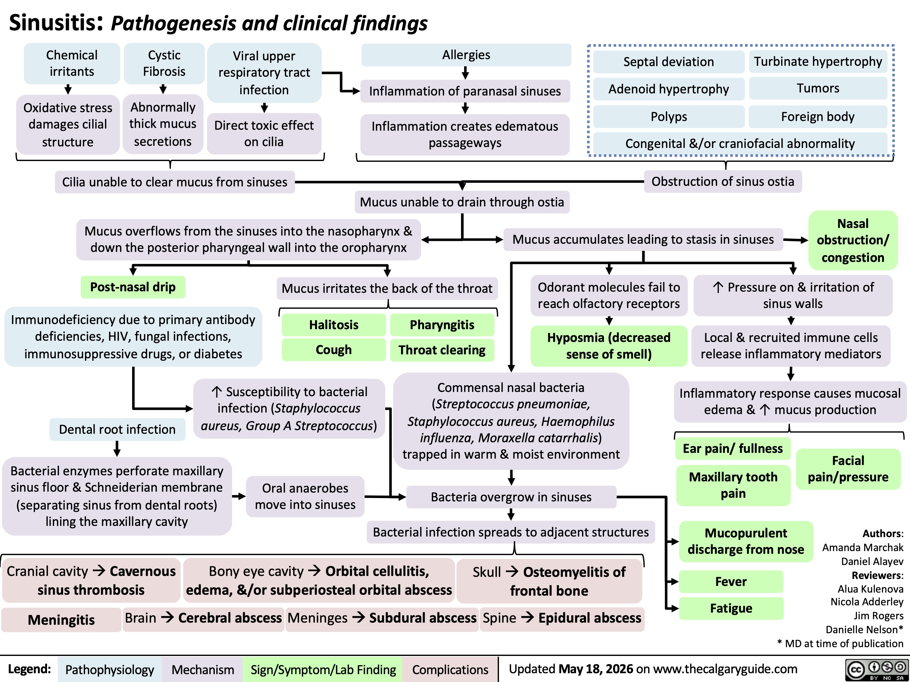
Sinusitis

Septal deviation Turbinate hypertrophy
Adenoid hypertrophy
Polyps
Tumors
Foreign body
Congenital &/or craniofacial abnormality
Sinusitis: Pathogenesis and clinical findings
Chemical
irritants
Cystic
Fibrosis
Viral upper
respiratory tract
infection
Allergies
Inflammation of paranasal sinuses
Oxidative stress
damages cilial
structure
Abnormally
thick mucus
secretions
Direct toxic effect
on cilia
Inflammation creates edematous
passageways
Cilia unable to clear mucus from sinuses
Obstruction of sinus ostia
Mucus unable to drain through ostia
Mucus overflows from the sinuses into the nasopharynx &
down the posterior pharyngeal wall into the oropharynx
Mucus accumulates leading to stasis in sinuses
Nasal
obstruction/
congestion
Post-nasal drip Mucus irritates the back of the throat
Odorant molecules fail to
reach olfactory receptors
↑ Pressure on & irritation of
sinus walls
Immunodeficiency due to primary antibody
deficiencies, HIV, fungal infections,
immunosuppressive drugs, or diabetes Cough
Halitosis Pharyngitis
Throat clearing
Hyposmia (decreased
sense of smell)
Local & recruited immune cells
release inflammatory mediators
↑ Susceptibility to bacterial
infection (Staphylococcus
Dental root infection
aureus, Group A Streptococcus)
Commensal nasal bacteria
(Streptococcus pneumoniae,
Staphylococcus aureus, Haemophilus
influenza, Moraxella catarrhalis)
trapped in warm & moist environment
Inflammatory response causes mucosal
edema & ↑ mucus production
Ear pain/ fullness
Bacterial enzymes perforate maxillary
sinus floor & Schneiderian membrane
(separating sinus from dental roots)
lining the maxillary cavity
Facial
pain/pressure
Oral anaerobes
move into sinuses
Bacteria overgrow in sinuses
Maxillary tooth
pain
Bacterial infection spreads to adjacent structures
Mucopurulent
discharge from nose
Cranial cavity à Cavernous
sinus thrombosis
Bony eye cavity à Orbital cellulitis,
edema, &/or subperiosteal orbital abscess
Skull à Osteomyelitis of
frontal bone
Fever
Fatigue
Brain à Cerebral abscess
Meninges à Subdural abscess
Meningitis Spine à Epidural abscess
Authors:
Amanda Marchak
Daniel Alayev
Reviewers:
Alua Kulenova
Nicola Adderley
Jim Rogers
Danielle Nelson*
* MD at time of publication
Updated May 18, 2026 on www.thecalgaryguide.com
Legend: Pathophysiology Mechanism
Sign/Symptom/Lab Finding Complications

© 2026 - The Calgary Guide to Understanding Disease

Disclaimer